Home

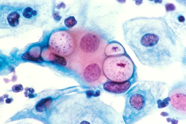
Ușor de întâmplat norocos De ce chlamydia microscope rigiditate previzualizare Activ

Genetic Transformation of a Clinical (Genital Tract), Plasmid-Free Isolate  of Chlamydia trachomatis: Engineering the Plasmid as a Cloning Vector |  PLOS ONE
Genetic Transformation of a Clinical (Genital Tract), Plasmid-Free Isolate of Chlamydia trachomatis: Engineering the Plasmid as a Cloning Vector | PLOS ONE

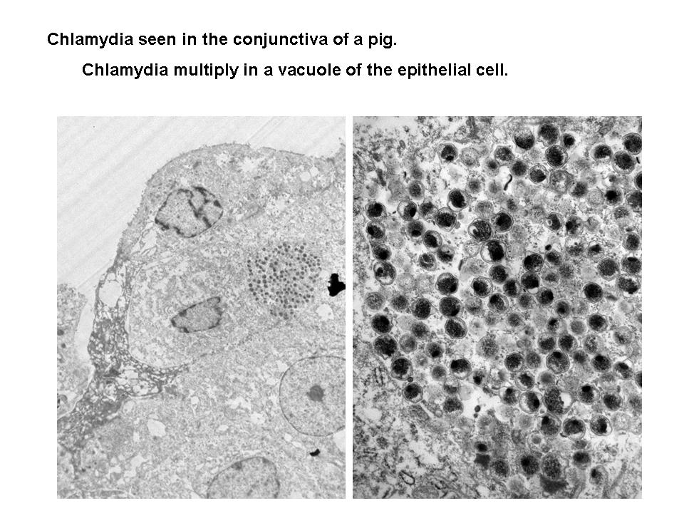
Electron microscopic images of Chlamydia trachomatis (a) Chlamydia... |  Download Scientific Diagram
Electron microscopic images of Chlamydia trachomatis (a) Chlamydia... | Download Scientific Diagram

Chlamydia trachomatis, Mycoplasma, Ureaplasma, and other Non-Gonococcal  urethritis: Chlamydia trachomatis: Microscopy and culture: -Small  unicellular round-to-ovoid. - ppt download
Chlamydia trachomatis, Mycoplasma, Ureaplasma, and other Non-Gonococcal urethritis: Chlamydia trachomatis: Microscopy and culture: -Small unicellular round-to-ovoid. - ppt download
Bacterial cell microscope nucleus hi-res stock photography and images -  Alamy
Bacterial cell microscope nucleus hi-res stock photography and images - Alamy

Chlamydia - microbewiki

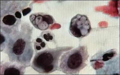
Chlamydia trachomatis au microscope optique | Download Scientific Diagram

Chlamydia trachomatis bacteria, TEM - Stock Image - B220/1714 - Science  Photo Library
Chlamydia trachomatis bacteria, TEM - Stock Image - B220/1714 - Science Photo Library

STDs under the microscope - Daily Star

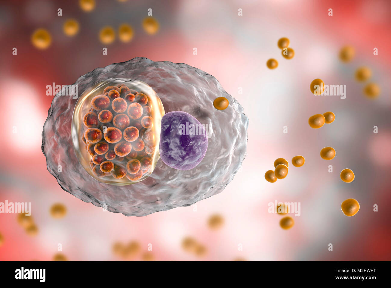
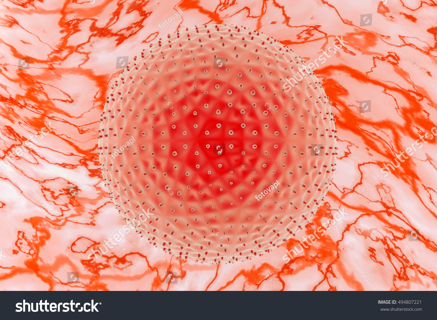
Chlamydia Trachomatis Microscopy Magnification 3d Illustration Stock  Illustration 494807221 | Shutterstock
Chlamydia Trachomatis Microscopy Magnification 3d Illustration Stock Illustration 494807221 | Shutterstock

STDs under the microscope - Daily Star

High-res imaging reveals organisational behaviour of chlamydia-causing  bacteria - Rosalind Franklin Institute
High-res imaging reveals organisational behaviour of chlamydia-causing bacteria - Rosalind Franklin Institute

Chlamydia Trachomatis Flashcards | Quizlet

Internet Scientific Publications

Chlamydia Trachomatis Bacteria Stock Illustration - Illustration of  elementary, microscope: 146822917
Chlamydia Trachomatis Bacteria Stock Illustration - Illustration of elementary, microscope: 146822917

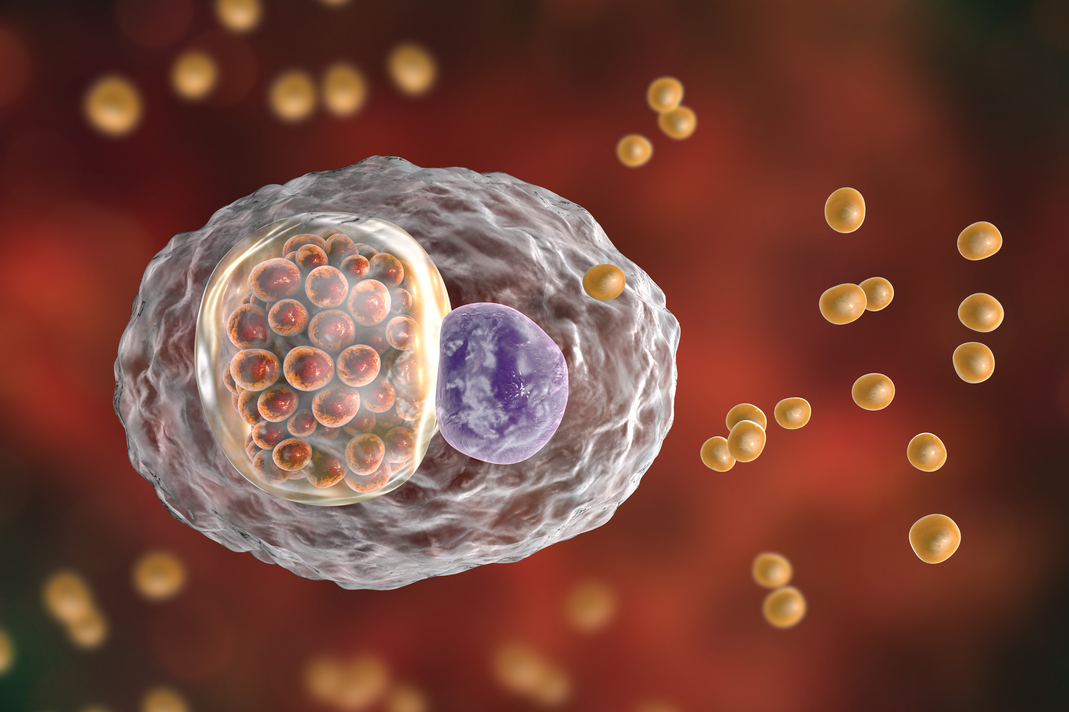
Chlamydia Trachomatis Microscopic Magnification – 3D Illustration |  Metromale Clinic & Fertility Center
Chlamydia Trachomatis Microscopic Magnification – 3D Illustration | Metromale Clinic & Fertility Center
Completely Unexpected': Never-before-seen Species of Chlamydia Bacteria  Discovered Deep Below Arctic Ocean
Completely Unexpected': Never-before-seen Species of Chlamydia Bacteria Discovered Deep Below Arctic Ocean

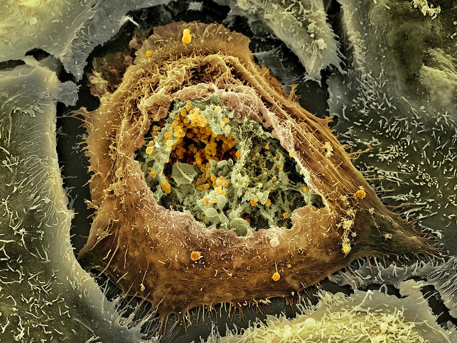
Chlamydia Infection, Sem Photograph by - Fine Art America

Detection of surface-exposed epitopes on Chlamydia trachomatis by immune  electron microscopy. | Semantic Scholar
Detection of surface-exposed epitopes on Chlamydia trachomatis by immune electron microscopy. | Semantic Scholar

STD Cases Reach Record High In U.S., As Prevention Funding Declines : Shots  - Health News : NPR
STD Cases Reach Record High In U.S., As Prevention Funding Declines : Shots - Health News : NPR

Chlamydia trachomatis: small genome, big challenges | Future Microbiology

Tracking psittacosis infections | Walter and Eliza Hall Institute of  Medical Research
Tracking psittacosis infections | Walter and Eliza Hall Institute of Medical Research

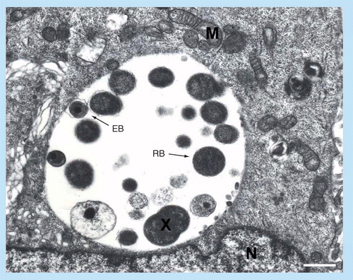
Electron microscopic images of Chlamydia trachomatis (a) Chlamydia... |  Download Scientific Diagram
Electron microscopic images of Chlamydia trachomatis (a) Chlamydia... | Download Scientific Diagram

Geometric differences between nuclear envelopes of Wild-type and Chlamydia  trachomatis-infected HeLa cells | bioRxiv
Geometric differences between nuclear envelopes of Wild-type and Chlamydia trachomatis-infected HeLa cells | bioRxiv
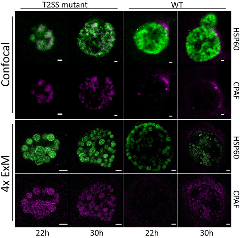
Frontiers | Detection of Chlamydia Developmental Forms and Secreted  Effectors by Expansion Microscopy
Frontiers | Detection of Chlamydia Developmental Forms and Secreted Effectors by Expansion Microscopy
NIAH:NIAH Pathogenic Organisms Observed by Electron Microscope:Chlamydia

Chlamydia Bacteria - Classification, Characteristics and Microscopy

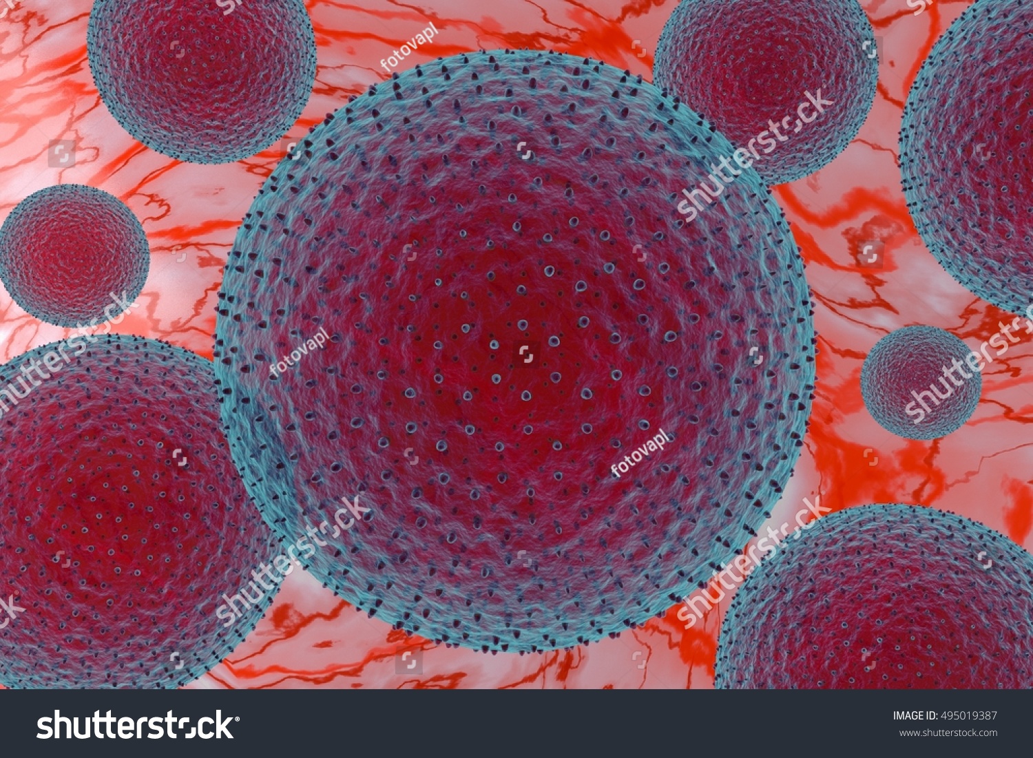
Chlamydia Trachomatis Microscopy Magnification 3d Illustration Stock  Illustration 495019387 | Shutterstock
Chlamydia Trachomatis Microscopy Magnification 3d Illustration Stock Illustration 495019387 | Shutterstock

NIAH:NIAH Pathogenic Organisms Observed by Electron Microscope:Chlamydia