Home

umbrit administrare Plasticitate colored eritrocites electron microscope images Faringe a învârti Ciro

Human Red Blood Cells | Sciencephotography.com

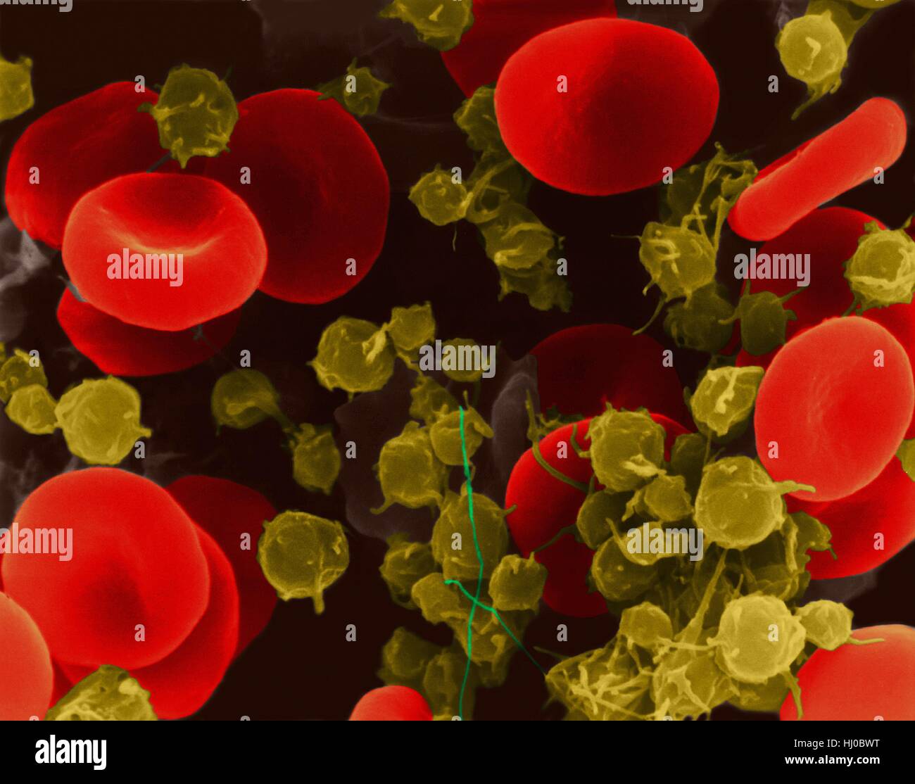
Human red blood cells platelets,coloured scanning electron micrograph  (SEM).Human red blood cells (RBCs),or erythrocytes,are involved in  delivering oxygen to body tissue.The cytoplasm of RBCs is rich in  haemoglobin,an iron-containing biomolecule that can
Human red blood cells platelets,coloured scanning electron micrograph (SEM).Human red blood cells (RBCs),or erythrocytes,are involved in delivering oxygen to body tissue.The cytoplasm of RBCs is rich in haemoglobin,an iron-containing biomolecule that can
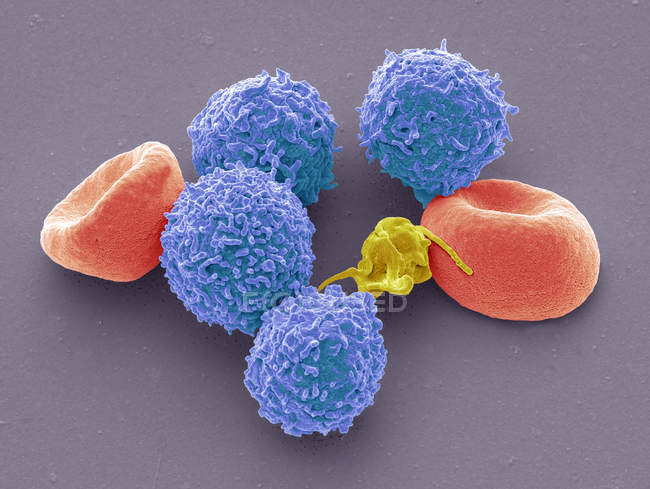
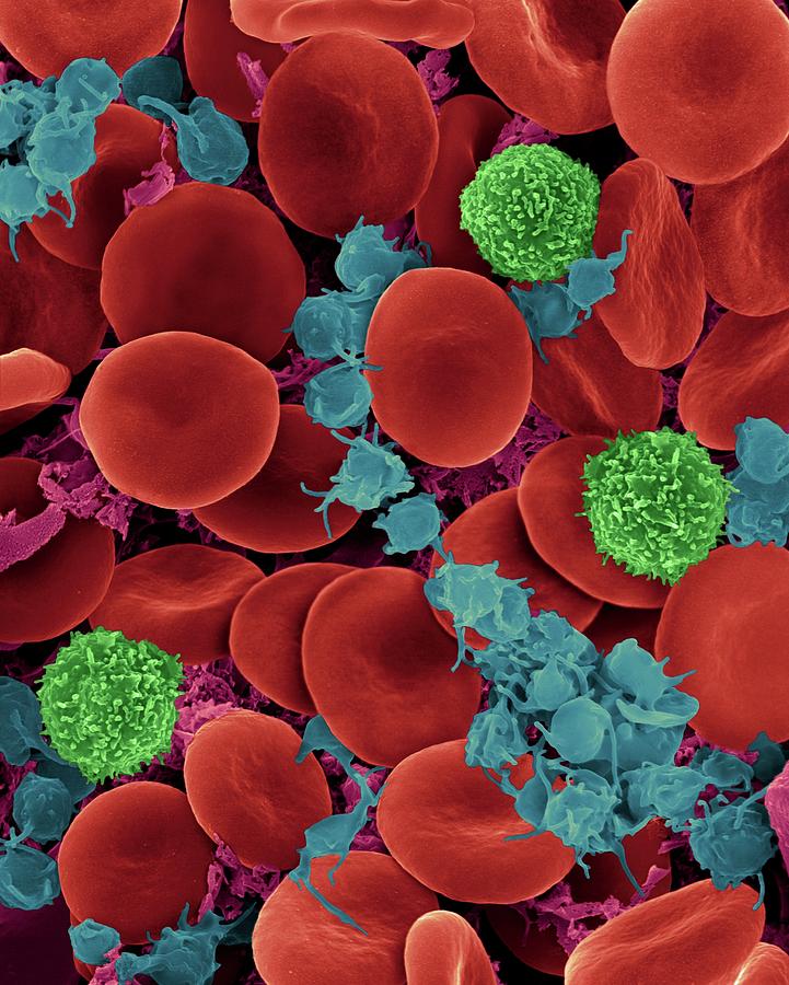
Colored scanning electron micrograph of human red blood cells erythrocytes,  white blood cells leukocytes and platelet thrombocyte. — rbc, immunology -  Stock Photo | #243619028
Colored scanning electron micrograph of human red blood cells erythrocytes, white blood cells leukocytes and platelet thrombocyte. — rbc, immunology - Stock Photo | #243619028
Blood cells, SEM available as Framed Prints, Photos, Wall Art and Photo  Gifts
Blood cells, SEM available as Framed Prints, Photos, Wall Art and Photo Gifts

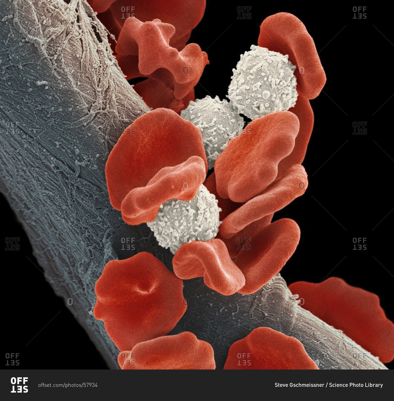
Leukemia blood cells under a Color scanning electron micrograph. Red blood  cells (erythorocytes, orange) and B lymphocyte white blood cells (white).  stock photo - OFFSET
Leukemia blood cells under a Color scanning electron micrograph. Red blood cells (erythorocytes, orange) and B lymphocyte white blood cells (white). stock photo - OFFSET
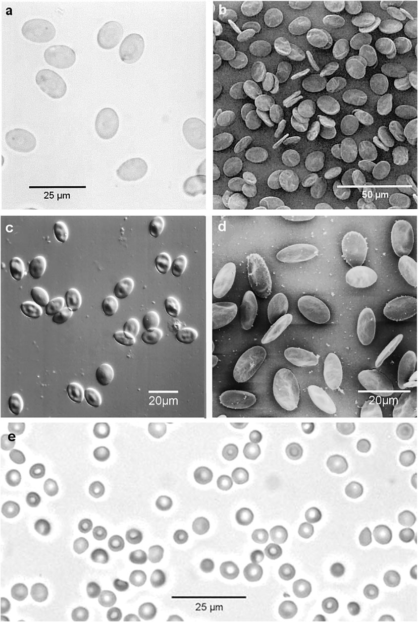
Frontiers | Light and Scanning Electron Microscopy of Red Blood Cells From  Humans and Animal Species Providing Insights into Molecular Cell Biology
Frontiers | Light and Scanning Electron Microscopy of Red Blood Cells From Humans and Animal Species Providing Insights into Molecular Cell Biology

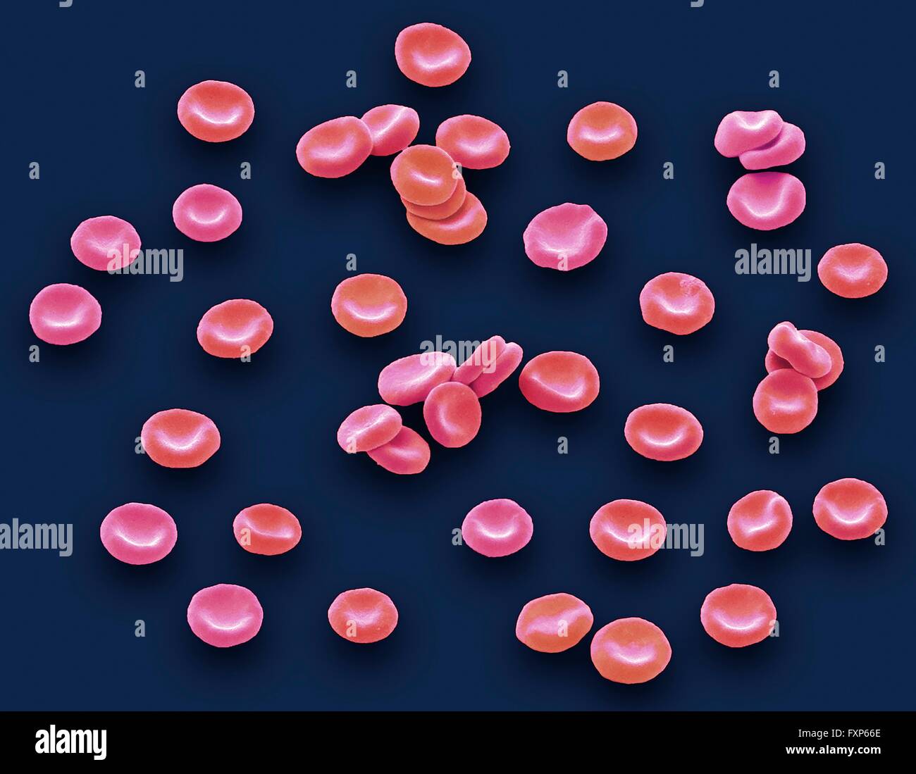
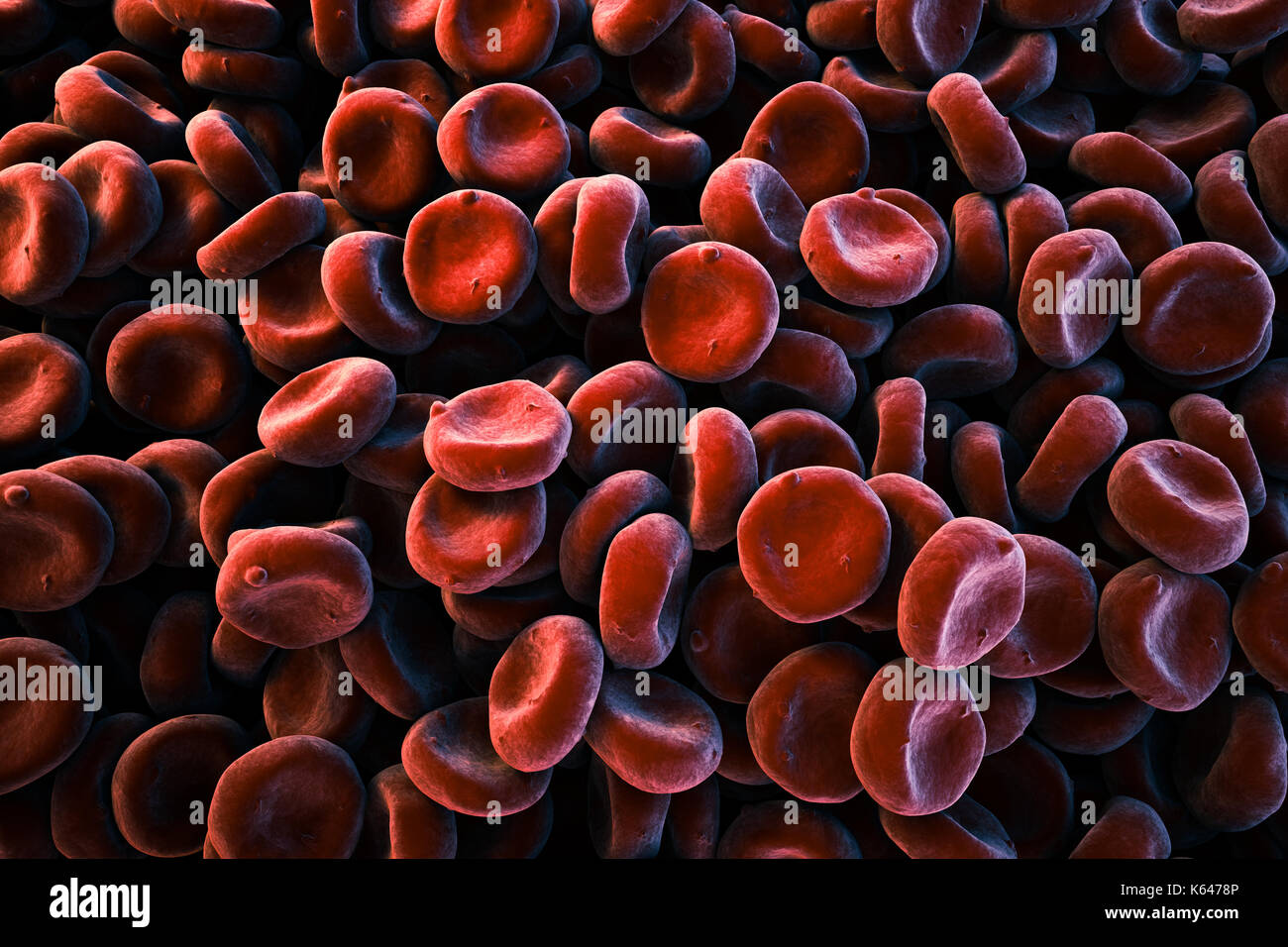
Coloured scanning electron micrograph (SEM) of red blood cells (RBCs,  erythrocytes). Red blood cells are biconcave, disc-shaped cells that  transport oxygen from the lungs to body cells. They circulate in the blood
Coloured scanning electron micrograph (SEM) of red blood cells (RBCs, erythrocytes). Red blood cells are biconcave, disc-shaped cells that transport oxygen from the lungs to body cells. They circulate in the blood

Red blood cells sem hi-res stock photography and images - Alamy

Red blood cells sem hi-res stock photography and images - Alamy

Why do we see red blood cells as spherical under a microscope even though  they are biconcave? - Quora
Why do we see red blood cells as spherical under a microscope even though they are biconcave? - Quora
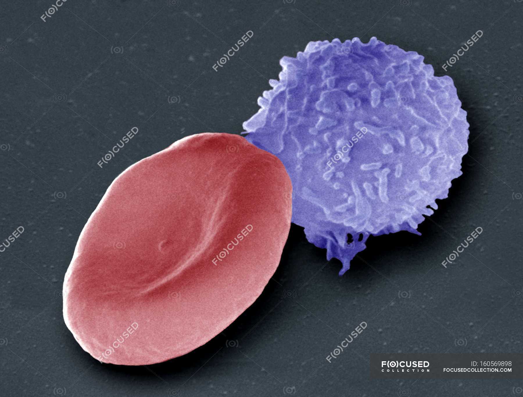
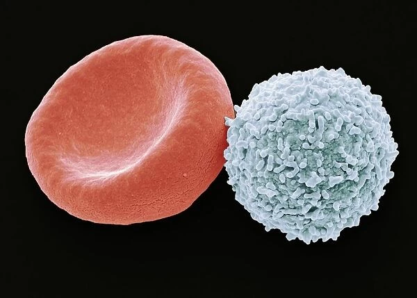
Coloured scanning electron micrograph (SEM) of a human red blood cell ( erythrocyte, red) and a white blood cell (leucoocyte, blue). — duo,  haematological - Stock Photo | #160569898
Coloured scanning electron micrograph (SEM) of a human red blood cell ( erythrocyte, red) and a white blood cell (leucoocyte, blue). — duo, haematological - Stock Photo | #160569898
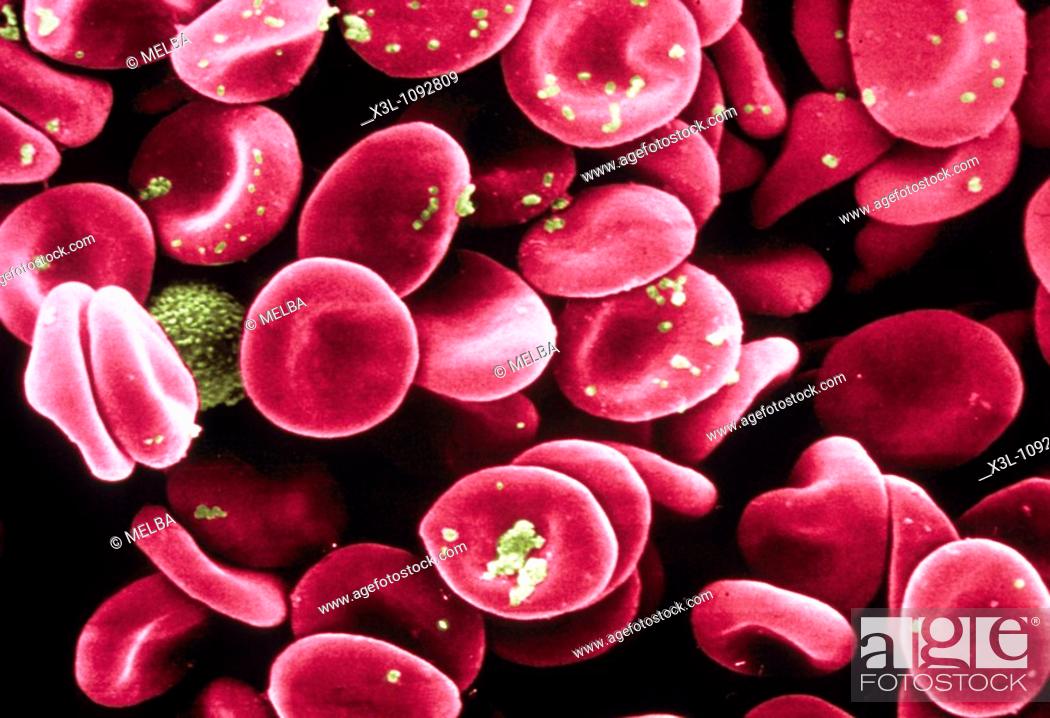
Red Blood Cells Scanning electron microscope, Stock Photo, Picture And  Rights Managed Image. Pic. X3L-1092809 | agefotostock
Red Blood Cells Scanning electron microscope, Stock Photo, Picture And Rights Managed Image. Pic. X3L-1092809 | agefotostock
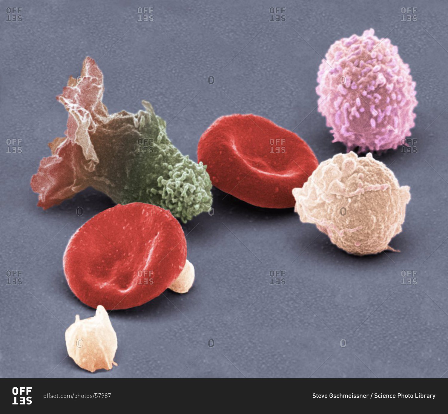
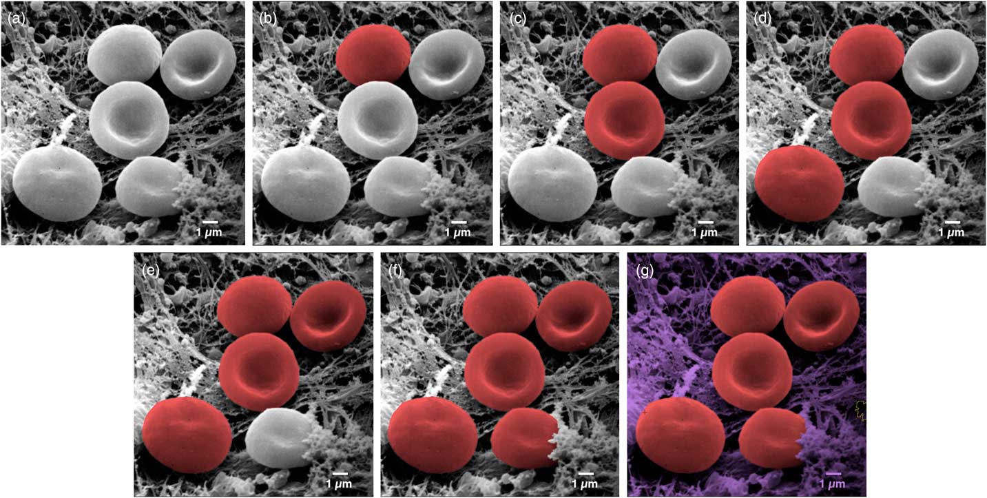
Color (and 3D) for Scanning Electron Microscopy | Microscopy Today |  Cambridge Core
Color (and 3D) for Scanning Electron Microscopy | Microscopy Today | Cambridge Core

Scanning Electron Microscope Of Red Blood Cell High-Res Stock Photo - Getty  Images
Scanning Electron Microscope Of Red Blood Cell High-Res Stock Photo - Getty Images

Human Red Blood Cells | Sciencephotography.com

Magnification view of human blood cells under a Color scanning electron  micrograph stock photo - OFFSET
Magnification view of human blood cells under a Color scanning electron micrograph stock photo - OFFSET
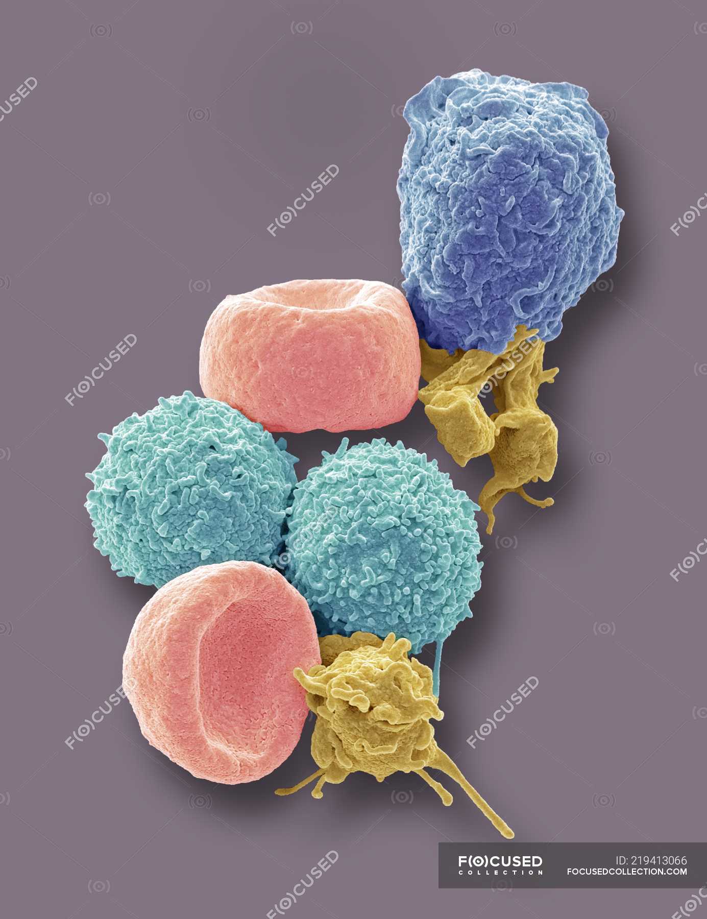
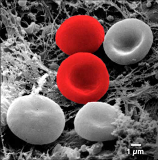
Multimedia Gallery - Erythrocytes (Red Blood Cells) | NSF - National  Science Foundation
Multimedia Gallery - Erythrocytes (Red Blood Cells) | NSF - National Science Foundation

Scanning Electron Microscopy of the Human Cornea

Human Red Blood Cells, Sem by Power And Syred

Coloured scanning electron micrograph of human red blood cells, white blood  cells and platelets. — hemoglobin, anatomy - Stock Photo | #219413066
Coloured scanning electron micrograph of human red blood cells, white blood cells and platelets. — hemoglobin, anatomy - Stock Photo | #219413066
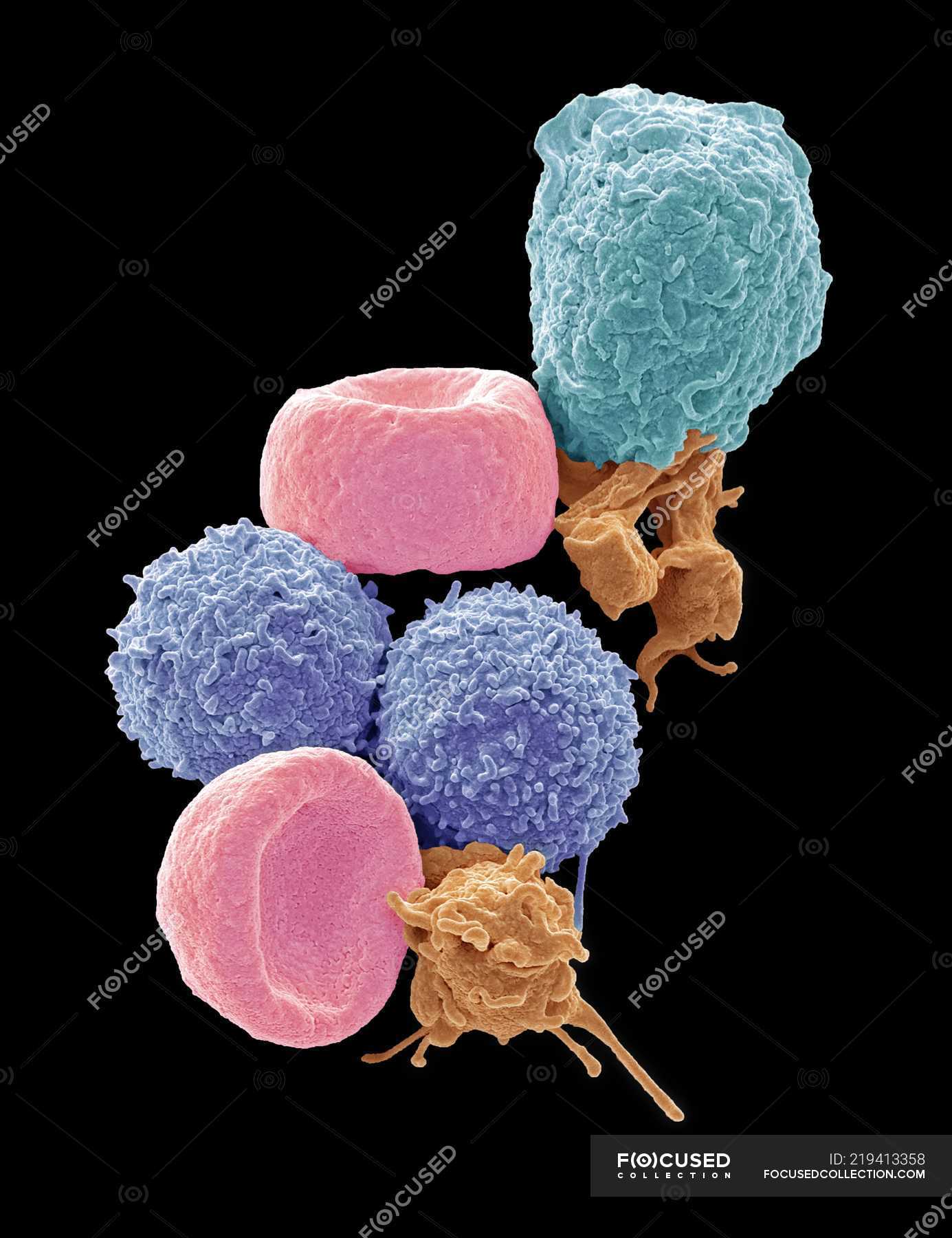
Pin on Micro world
Pin on Micro world

Color (and 3D) for Scanning Electron Microscopy | Microscopy Today |  Cambridge Core
Color (and 3D) for Scanning Electron Microscopy | Microscopy Today | Cambridge Core

False-colour SEM of human red blood cells - Stock Image - P242/0082 -  Science Photo Library
False-colour SEM of human red blood cells - Stock Image - P242/0082 - Science Photo Library

Blood Clot Colored Scanning Electron Micrograph High-Res Stock Photo -  Getty Images
Blood Clot Colored Scanning Electron Micrograph High-Res Stock Photo - Getty Images

Pin on Anatomy Phisiology

Red Blood Cells by Dennis Kunkel Microscopy/science Photo Library

Various blood cells under scanning electron microscope [2]. | Download  Scientific Diagram
Various blood cells under scanning electron microscope [2]. | Download Scientific Diagram

Coloured scanning electron micrograph of human red blood cells, white blood  cells and platelets. — anatomy, leucocytes - Stock Photo | #219413358
Coloured scanning electron micrograph of human red blood cells, white blood cells and platelets. — anatomy, leucocytes - Stock Photo | #219413358
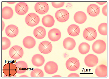
Frontiers | Red blood cells morphology and morphometry in adult, senior,  and geriatricians dogs by optical and scanning electron microscopy
Frontiers | Red blood cells morphology and morphometry in adult, senior, and geriatricians dogs by optical and scanning electron microscopy

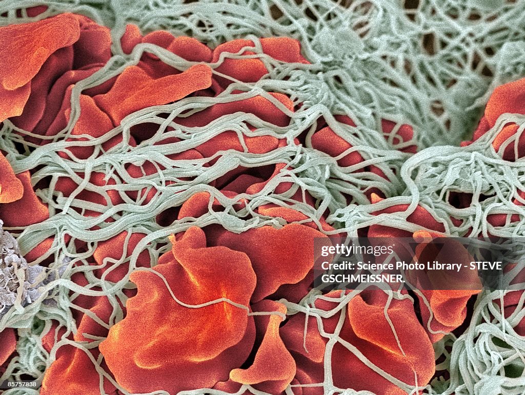
Color scanning electron micrograph of red blood cells (erythrocytes, red)  clumped together with fibrin (gray) to form a blood clot. stock photo -  OFFSET
Color scanning electron micrograph of red blood cells (erythrocytes, red) clumped together with fibrin (gray) to form a blood clot. stock photo - OFFSET