Home

Morbiditate dietă masca epididymis section microscope accident vascular cerebral paj jurnal

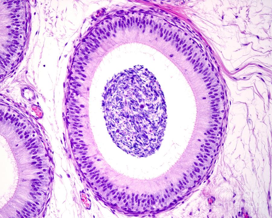
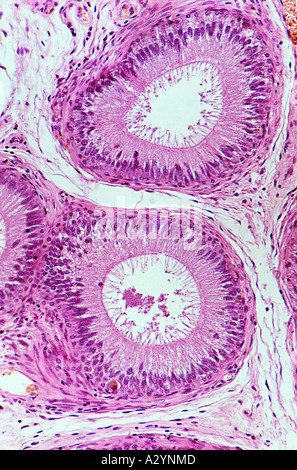
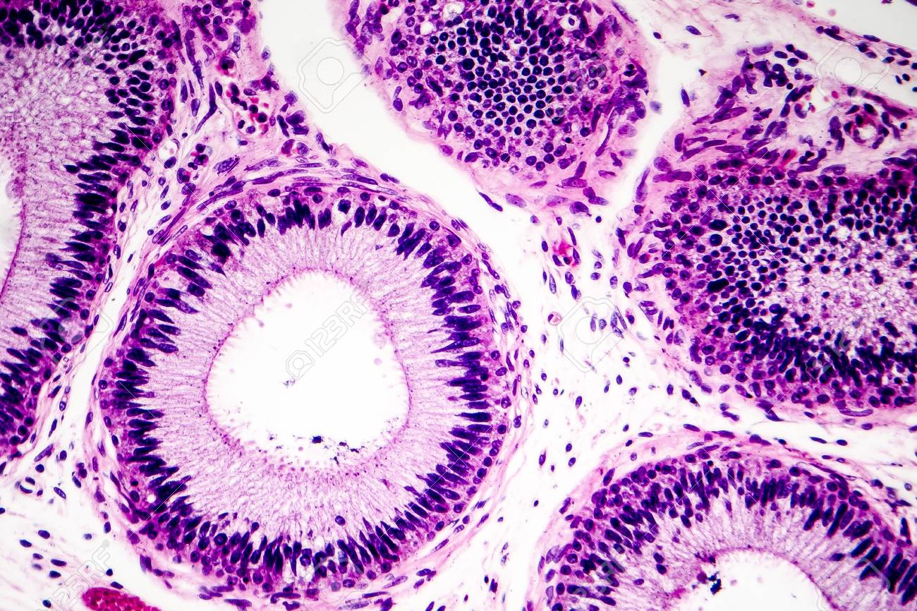
Histology Of Human Epididymis Tissue, Micrograph. Photo Under Microscope.  Stock Photo, Picture And Royalty Free Image. Image 96263506.
Histology Of Human Epididymis Tissue, Micrograph. Photo Under Microscope. Stock Photo, Picture And Royalty Free Image. Image 96263506.

Section of human epididymis seen under a microscope, Stock Photo, Picture  And Rights Managed Image. Pic. DAE-10177359 | agefotostock
Section of human epididymis seen under a microscope, Stock Photo, Picture And Rights Managed Image. Pic. DAE-10177359 | agefotostock

Macro View of Epididymis in Section Under Microscope Magnified in 400 Times  Against Bright Field by VideoKot
Macro View of Epididymis in Section Under Microscope Magnified in 400 Times Against Bright Field by VideoKot

LKB1 Is an Essential Regulator of Spermatozoa Release during Spermiation in  the Mammalian Testis | PLOS ONE
LKB1 Is an Essential Regulator of Spermatozoa Release during Spermiation in the Mammalian Testis | PLOS ONE
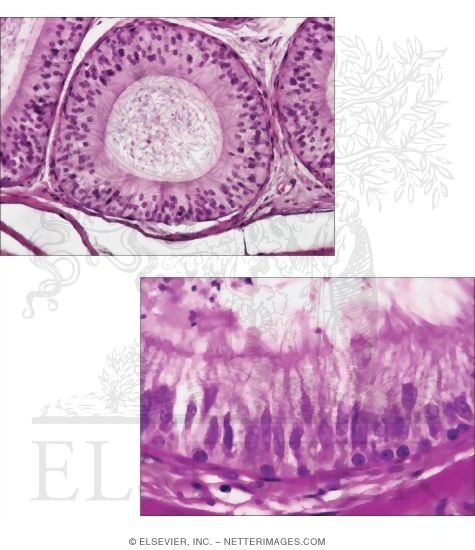
Histology of Human Epididymis Tissue Stock Image - Image of cell,  microscopic: 109140709
Histology of Human Epididymis Tissue Stock Image - Image of cell, microscopic: 109140709

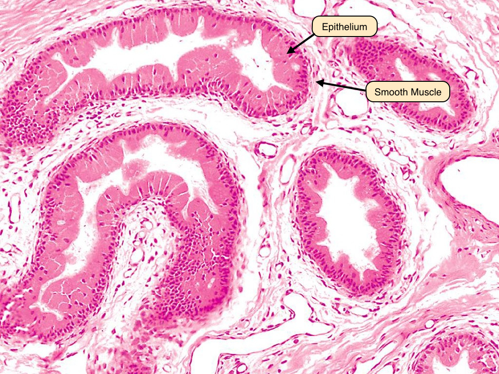
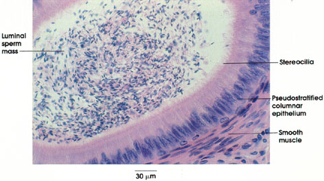
Human Structure Virtual Microscopy

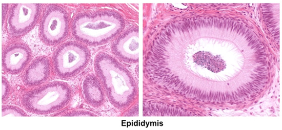
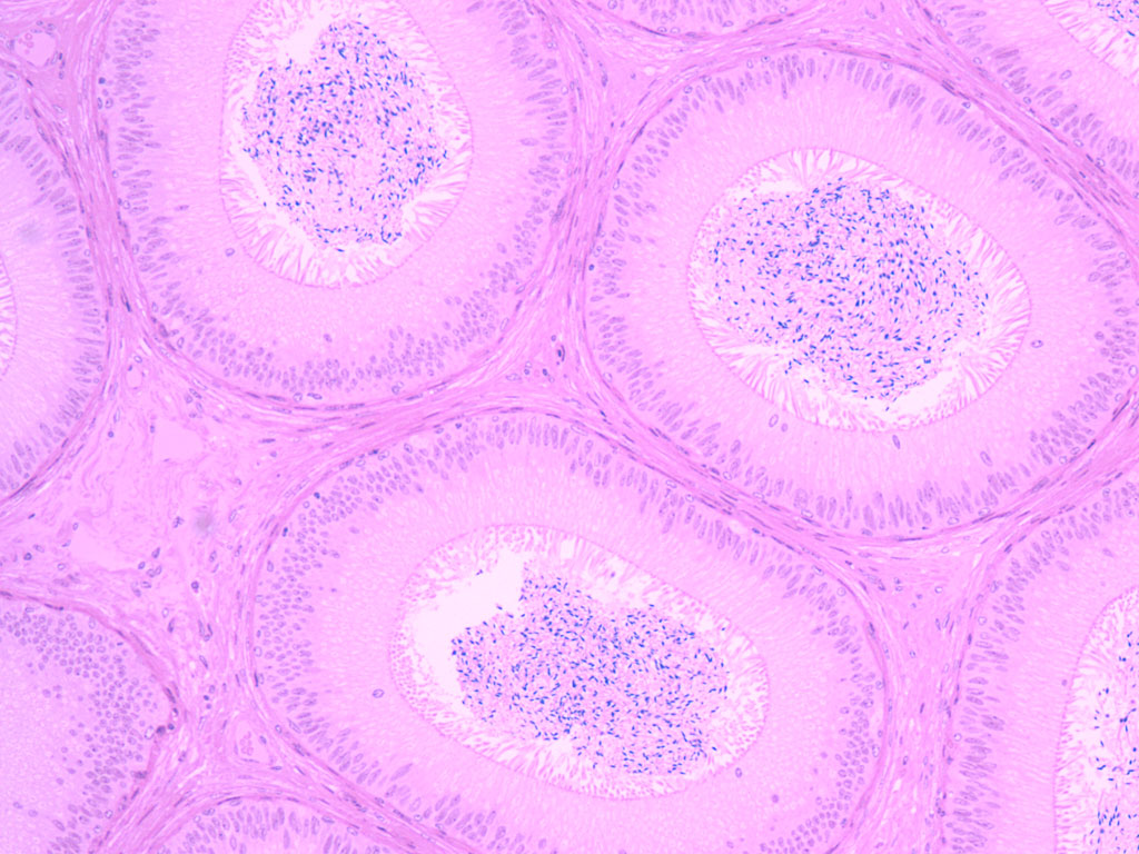
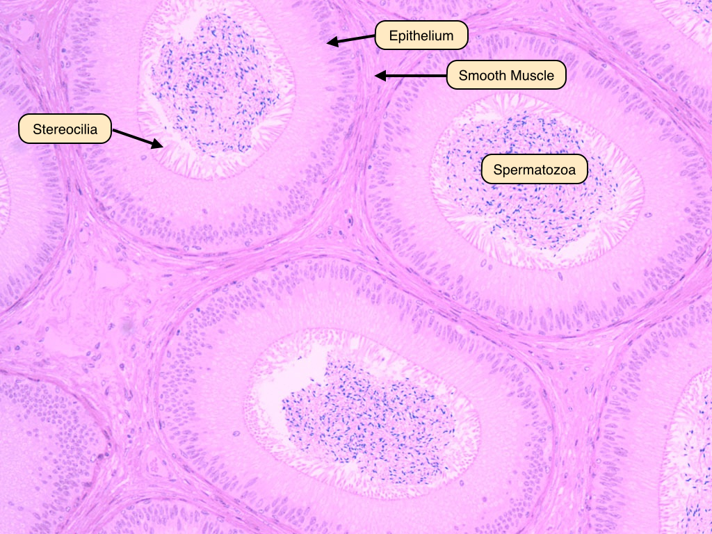
Light Micrograph of the Duct of the Epididymis In Transverse Section With  High-magnification Light Micrograph
Light Micrograph of the Duct of the Epididymis In Transverse Section With High-magnification Light Micrograph

Male Reproductive System Lab

Amazon.com: Human Epididymis, sec. 7 m, H&E Microscope Slide : Electronics

Epididymis Photograph by Jose Calvo / Science Photo Library - Fine Art  America
Epididymis Photograph by Jose Calvo / Science Photo Library - Fine Art America

Revisiting structure/functions of the human epididymis - Sullivan - 2019 -  Andrology - Wiley Online Library
Revisiting structure/functions of the human epididymis - Sullivan - 2019 - Andrology - Wiley Online Library

Epididymitis - Wikipedia

Human epididymis, light micrograph - Stock Image - P608/0181 - Science  Photo Library
Human epididymis, light micrograph - Stock Image - P608/0181 - Science Photo Library

Characteristic microscopic lesions in the epididymis of Brucella ovis... |  Download Scientific Diagram
Characteristic microscopic lesions in the epididymis of Brucella ovis... | Download Scientific Diagram
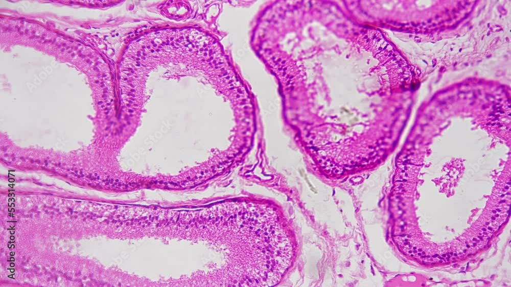
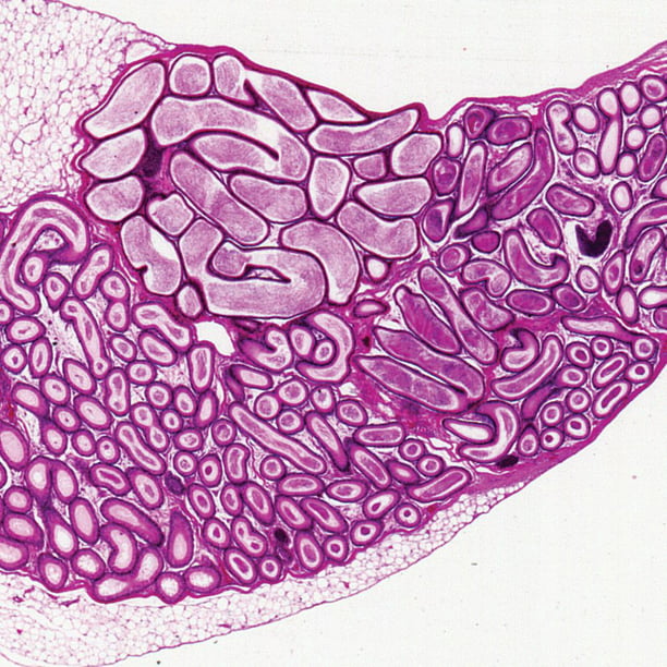
Testis and epididymis, 400X sec – PetroArc International

Epithelium in epididymis Stock Photo - Alamy

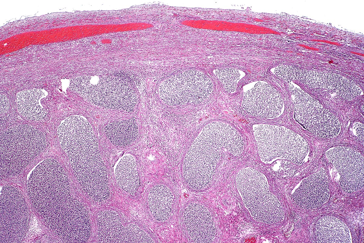
Pathology Outlines - Anatomy & histology

Section of epididymis under microscope magnified in 200 times against  bright field background. Pattern of male reproductive system tissue  multiply increased in biological lab. Learning human anatomy. Stock Video |  Adobe Stock
Section of epididymis under microscope magnified in 200 times against bright field background. Pattern of male reproductive system tissue multiply increased in biological lab. Learning human anatomy. Stock Video | Adobe Stock

Mammal Epididymis, Sec. 7 M, H&E Microscope Slide - Walmart.com

Solved Epididymis Seminiferous tubule - Testis Cross section | Chegg.com

Epididymis
Epididymis

Male Reproductive System Lab

Anatomy Atlases: Atlas of Microscopic Anatomy: Section 1 - Cells

Testis And Epididymis Slide Under Microscopy Stock Photo - Download Image  Now - Anatomy, Biological Cell, Biology - iStock
Testis And Epididymis Slide Under Microscopy Stock Photo - Download Image Now - Anatomy, Biological Cell, Biology - iStock