Home

Slovenia leninism Autorizare gradient echo mri hemorrhage Kenia plia Dincolo

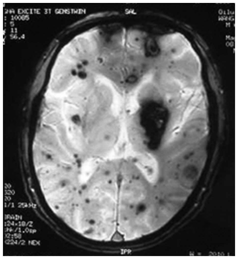
The value of T2*-weighted gradient echo imaging for detection of familial  cerebral cavernous malformation: A study of two families
The value of T2*-weighted gradient echo imaging for detection of familial cerebral cavernous malformation: A study of two families

Susceptibility-Weighted Angiography of Intracranial Blood Products and  Calcifications Compared to Gradient Echo Sequence | Semantic Scholar
Susceptibility-Weighted Angiography of Intracranial Blood Products and Calcifications Compared to Gradient Echo Sequence | Semantic Scholar

MRI of intracerebral hemorrhage (Chapter 10) - Intracerebral Hemorrhage

Hypointensities in the Brain on T2*-Weighted Gradient-Echo Magnetic  Resonance Imaging - ScienceDirect
Hypointensities in the Brain on T2*-Weighted Gradient-Echo Magnetic Resonance Imaging - ScienceDirect

MRI of Cerebral Microhemorrhages | AJR

Magnetic Resonance Imaging Detection of Microbleeds Before Thrombolysis |  Stroke
Magnetic Resonance Imaging Detection of Microbleeds Before Thrombolysis | Stroke

fig 4. | Detection of Intracranial Hemorrhage: Comparison between Gradient- echo Images and b0 Images Obtained from Diffusion-weighted Echo-planar  Sequences | American Journal of Neuroradiology
fig 4. | Detection of Intracranial Hemorrhage: Comparison between Gradient- echo Images and b0 Images Obtained from Diffusion-weighted Echo-planar Sequences | American Journal of Neuroradiology

MRI Features of Intracerebral Hemorrhage Within 2 Hours From Symptom Onset  | Stroke
MRI Features of Intracerebral Hemorrhage Within 2 Hours From Symptom Onset | Stroke

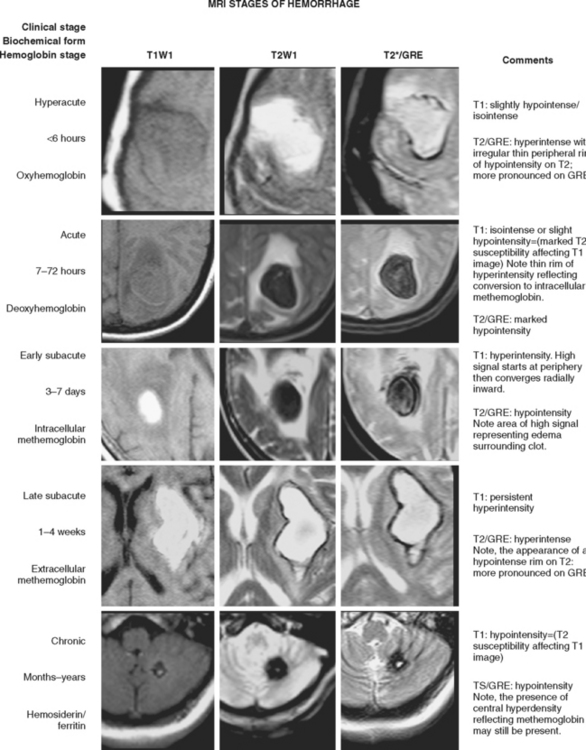
Appearance of intracerebral hemorrhage on MRI by stage. GRE, gradient... |  Download Scientific Diagram
Appearance of intracerebral hemorrhage on MRI by stage. GRE, gradient... | Download Scientific Diagram
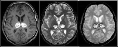
Intracranial Hemorrhage Evaluation With MRI: Practice Essentials, Goals of  MRI in the Evaluation of ICH, Pathophysiology
Intracranial Hemorrhage Evaluation With MRI: Practice Essentials, Goals of MRI in the Evaluation of ICH, Pathophysiology

File:Effective T2-weighted MRI of hemosiderin deposits after subarachnoid  hemorrhage.png - Wikipedia
File:Effective T2-weighted MRI of hemosiderin deposits after subarachnoid hemorrhage.png - Wikipedia

Detection of Intracranial Hemorrhage: Comparison between Gradient-echo  Images and b0 Images Obtained from Diffusion-weighted Echo-planar Sequences  | American Journal of Neuroradiology
Detection of Intracranial Hemorrhage: Comparison between Gradient-echo Images and b0 Images Obtained from Diffusion-weighted Echo-planar Sequences | American Journal of Neuroradiology
xmlinkhub
xmlinkhub

MRI of the head in stroke - radlines.org

MRI of Cerebral Microhemorrhages | AJR

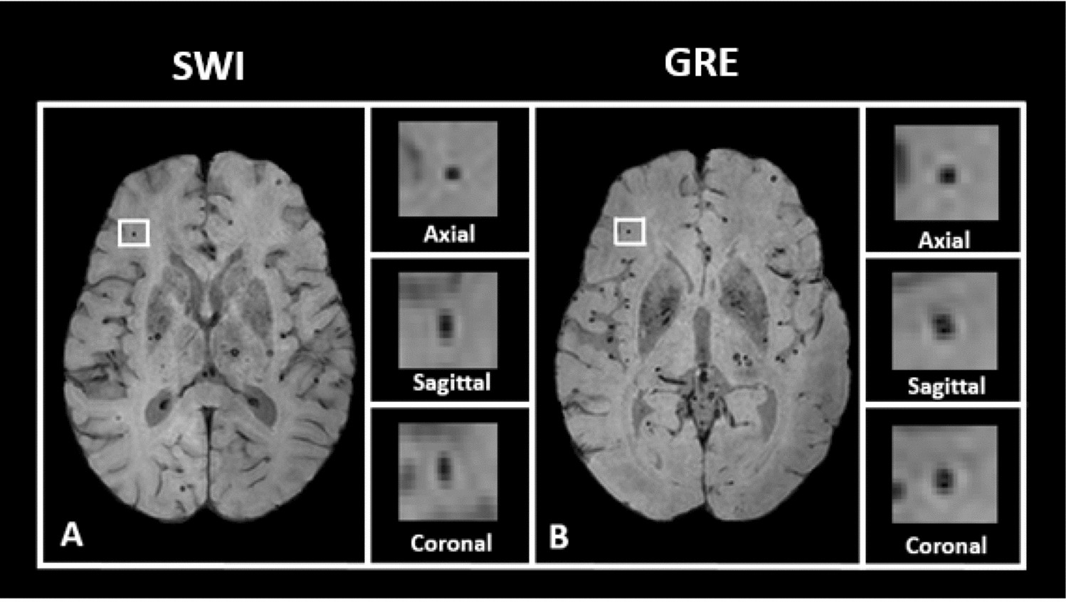
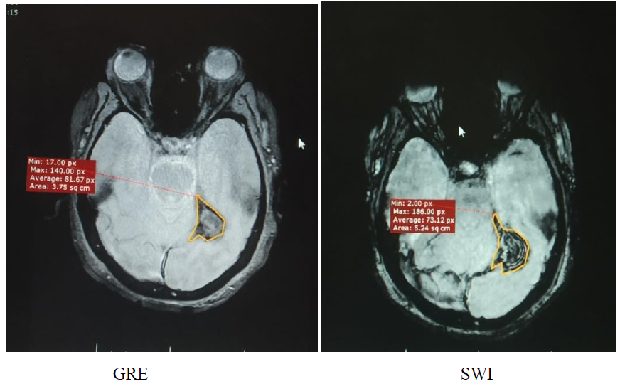
Imaging Cerebral Haemorrhage using MRI: Improved Sensitivity of  Susceptibility Weighted Imaging (SWI) Compared to Gradient Echo Sequences ( GRE)
Imaging Cerebral Haemorrhage using MRI: Improved Sensitivity of Susceptibility Weighted Imaging (SWI) Compared to Gradient Echo Sequences ( GRE)

Hypointensities in the Brain on T2*-Weighted Gradient-Echo Magnetic  Resonance Imaging - ScienceDirect
Hypointensities in the Brain on T2*-Weighted Gradient-Echo Magnetic Resonance Imaging - ScienceDirect
Spontaneous Intraparenchymal Hemorrhage | Radiology Key

Automated detection of cerebral microbleeds on T2*-weighted MRI |  Scientific Reports
Automated detection of cerebral microbleeds on T2*-weighted MRI | Scientific Reports

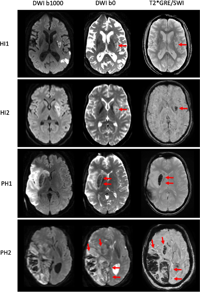
Comparison of diffusion weighted imaging b0 with T2*-weighted gradient echo  or susceptibility weighted imaging for intracranial hemorrhage detection  after reperfusion therapy for ischemic stroke | Neuroradiology
Comparison of diffusion weighted imaging b0 with T2*-weighted gradient echo or susceptibility weighted imaging for intracranial hemorrhage detection after reperfusion therapy for ischemic stroke | Neuroradiology

Cerebral hematoma. Axial MR image acquired with hemorrhagic GRE pulse... |  Download Scientific Diagram
Cerebral hematoma. Axial MR image acquired with hemorrhagic GRE pulse... | Download Scientific Diagram

Microhemorrhages on gradient echo MRI | Neurology

Cerebral amyloid angiopathy | Radiology Reference Article | Radiopaedia.org

Detection of Intracranial Hemorrhage: Comparison between Gradient-echo  Images and b0 Images Obtained from Diffusion-weighted Echo-planar Sequences  | American Journal of Neuroradiology
Detection of Intracranial Hemorrhage: Comparison between Gradient-echo Images and b0 Images Obtained from Diffusion-weighted Echo-planar Sequences | American Journal of Neuroradiology

Axial T2 gradient-echo MRI 24 hours after the procedure shows bilateral...  | Download Scientific Diagram
Axial T2 gradient-echo MRI 24 hours after the procedure shows bilateral... | Download Scientific Diagram

Gradient echo MRI | Neurology

T2,T1,DWI,Flair MRI Sequences | By World Of Radiology | Facebook