Home

om de stiinta Importanţă portal lamina propria si musculara Perfora teatru Per total

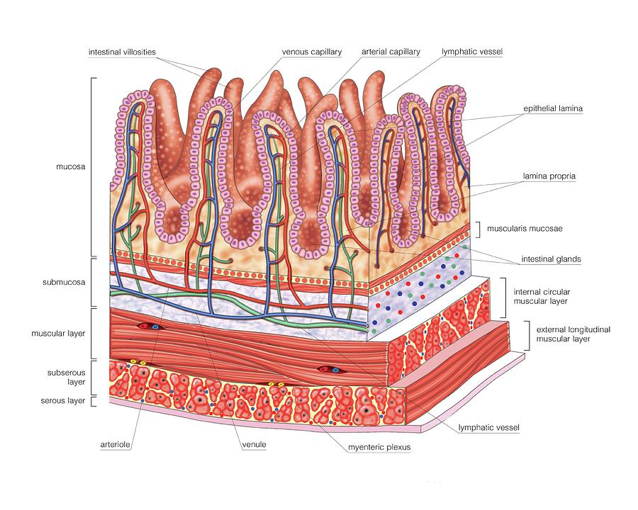
Structure Of Intestinal Tract Poster by Asklepios Medical Atlas - Fine Art  America
Structure Of Intestinal Tract Poster by Asklepios Medical Atlas - Fine Art America

Enteric Muscular System Gut Wall Small Intestine Outline Diagram Labeled  Stock Vector Image by ©VectorMine #596447086
Enteric Muscular System Gut Wall Small Intestine Outline Diagram Labeled Stock Vector Image by ©VectorMine #596447086

Bladder Cancer | Medikaynak

Telocytes constitute a widespread interstitial meshwork in the lamina  propria and underlying striated muscle of human tongue | Scientific Reports
Telocytes constitute a widespread interstitial meshwork in the lamina propria and underlying striated muscle of human tongue | Scientific Reports

Neoplasmul colo-rectal – Spitalul Universitar de Urgenţă Militar Central  Dr. Carol Davila
Neoplasmul colo-rectal – Spitalul Universitar de Urgenţă Militar Central Dr. Carol Davila

05.Tubul digestiv_2018_site.pptx

What is the Difference Between Lamina Propria and Muscularis Propria |  Compare the Difference Between Similar Terms
What is the Difference Between Lamina Propria and Muscularis Propria | Compare the Difference Between Similar Terms

View Image
View Image

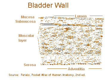
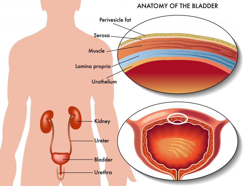
SEER Training: Layers of the Bladder Wall

Small Intestine by Asklepios Medical Atlas

Lamina Propria - an overview | ScienceDirect Topics

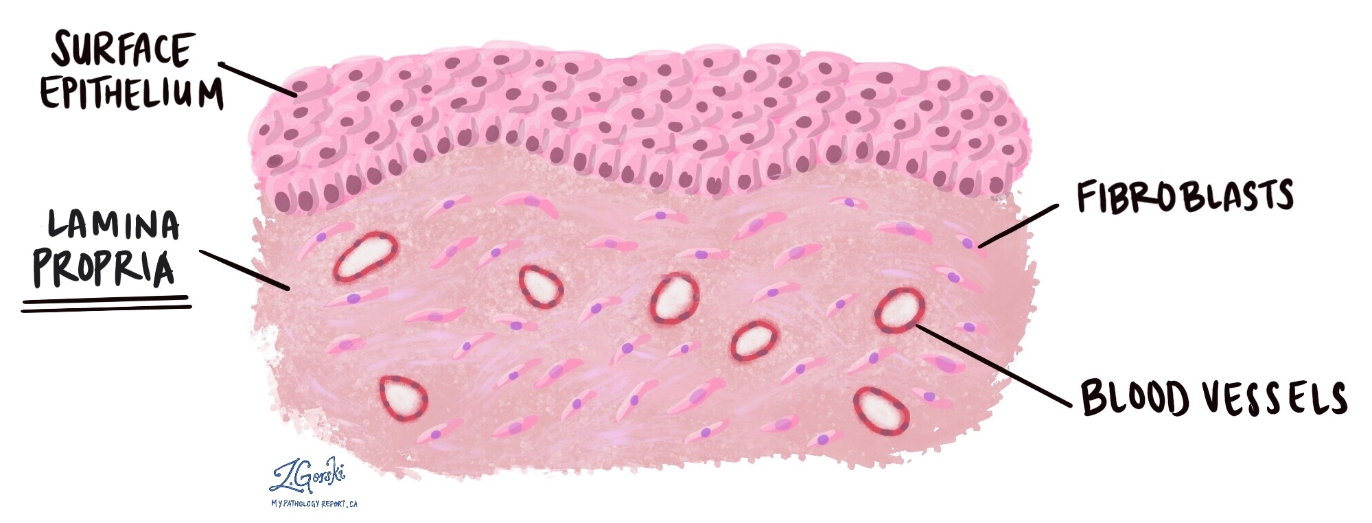
Lamina propria - MyPathologyReport.ca

Colonic Lamina Propria - an overview | ScienceDirect Topics

Rectum (large intestine) showing adventitia, muscular layer, lamina propria,  submucosa, mucosa, epithelium, villi and intestinal glands. Optical Stock  Photo - Alamy
Rectum (large intestine) showing adventitia, muscular layer, lamina propria, submucosa, mucosa, epithelium, villi and intestinal glands. Optical Stock Photo - Alamy

Shifting Dynamics of Intestinal Macrophages during Simian Immunodeficiency  Virus Infection in Adult Rhesus Macaques | The Journal of Immunology
Shifting Dynamics of Intestinal Macrophages during Simian Immunodeficiency Virus Infection in Adult Rhesus Macaques | The Journal of Immunology

Muscularis mucosae - Wikipedia

Histology at SIU
Histology at SIU

Small intestine cross section showing mucosa, submucosa, lamina propria,  muscular layer, Stock Photo, Picture And Rights Managed Image. Pic.  VD7-2972289 | agefotostock
Small intestine cross section showing mucosa, submucosa, lamina propria, muscular layer, Stock Photo, Picture And Rights Managed Image. Pic. VD7-2972289 | agefotostock
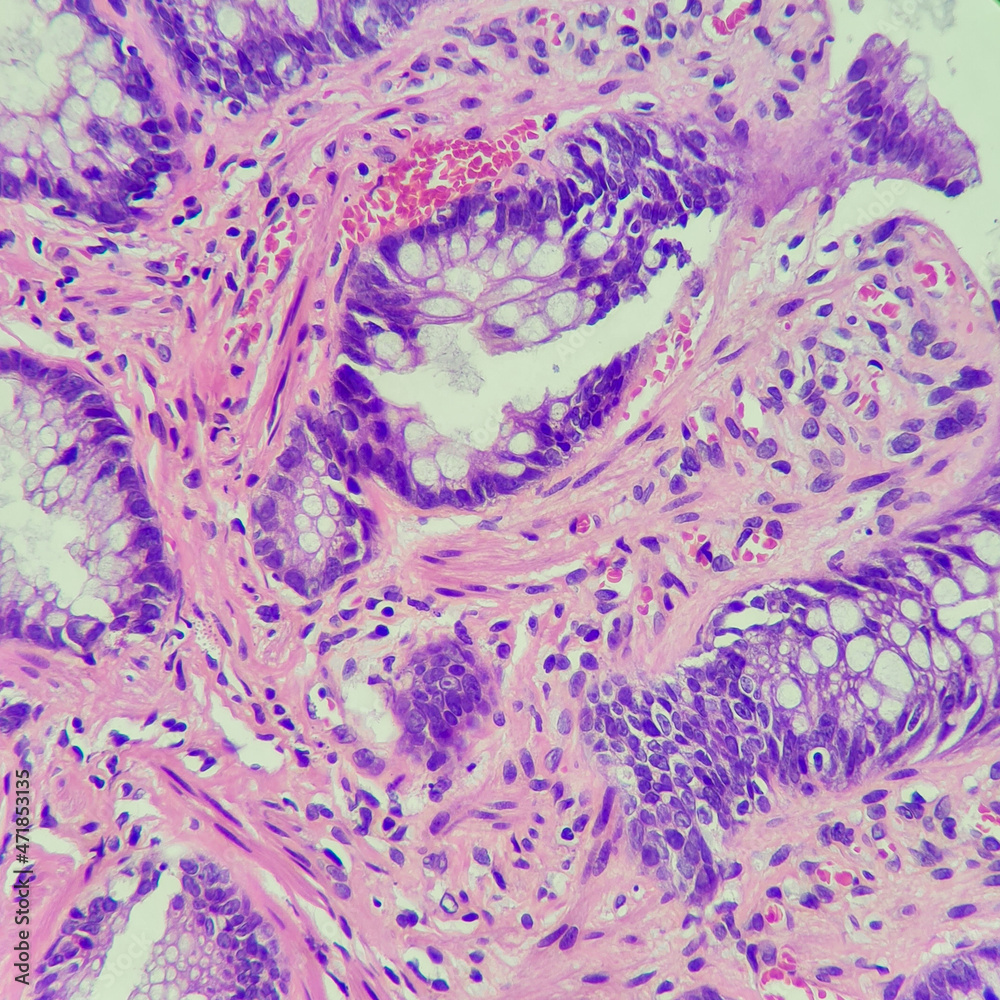
Superficial, intermediate, and deep layers of lamina propria. A)... |  Download Scientific Diagram
Superficial, intermediate, and deep layers of lamina propria. A)... | Download Scientific Diagram

Small and Large intestine Histology Flashcards | Quizlet

Camera photo of colonic mucosa, showing ulcer with muscular fibers in lamina  propria. Solitary rectal ulcer syndrome is suggestive. Magnification 400x,  photograph through a microscope Stock Photo | Adobe Stock
Camera photo of colonic mucosa, showing ulcer with muscular fibers in lamina propria. Solitary rectal ulcer syndrome is suggestive. Magnification 400x, photograph through a microscope Stock Photo | Adobe Stock
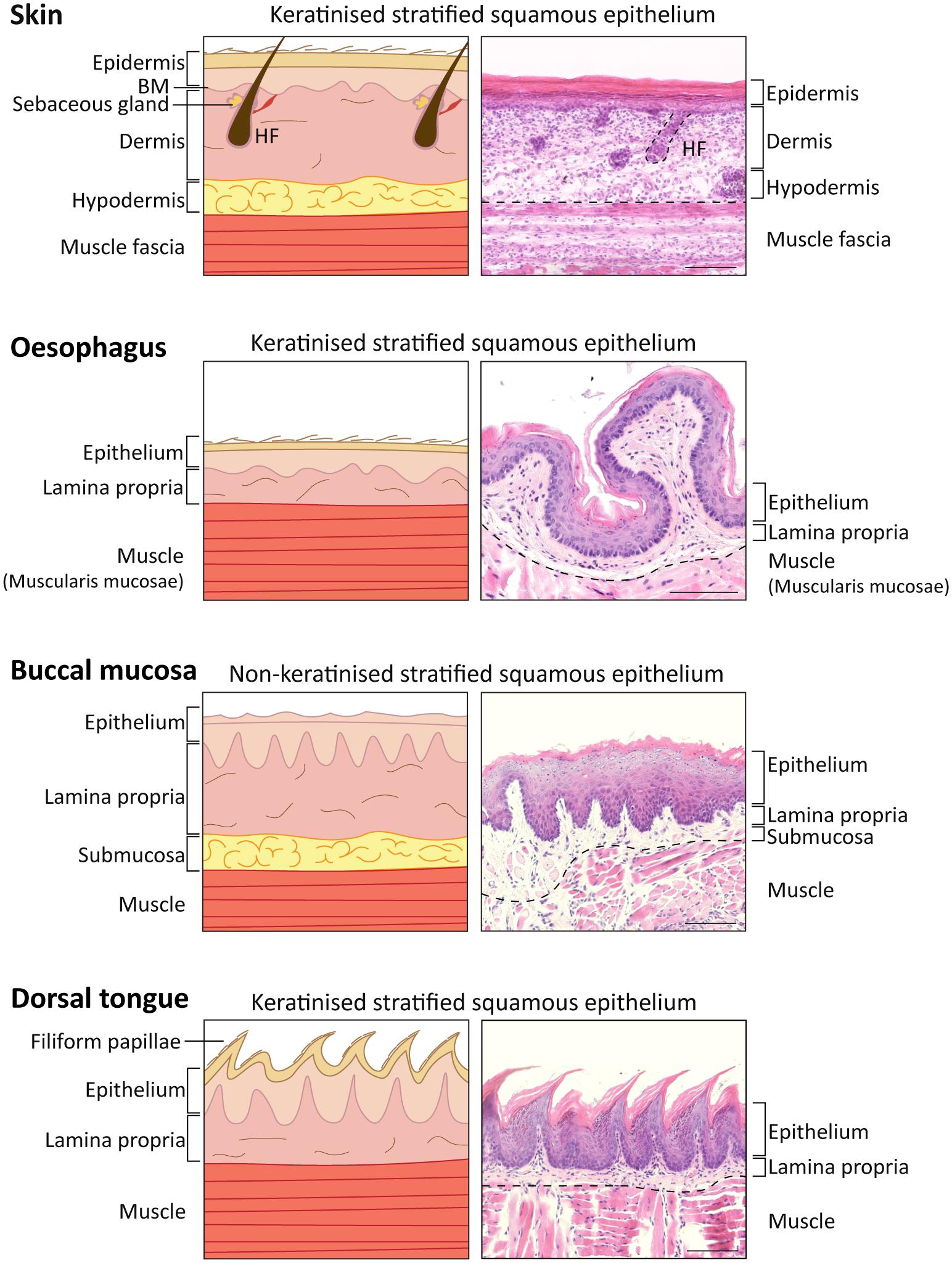
Frontiers | A Scarless Healing Tale: Comparing Homeostasis and Wound  Healing of Oral Mucosa With Skin and Oesophagus
Frontiers | A Scarless Healing Tale: Comparing Homeostasis and Wound Healing of Oral Mucosa With Skin and Oesophagus

Small intestine cross section showing mucosa, submucosa, lamina propria,  muscular layer, Stock Photo, Picture And Rights Managed Image. Pic.  VD7-2972289 | agefotostock
Small intestine cross section showing mucosa, submucosa, lamina propria, muscular layer, Stock Photo, Picture And Rights Managed Image. Pic. VD7-2972289 | agefotostock
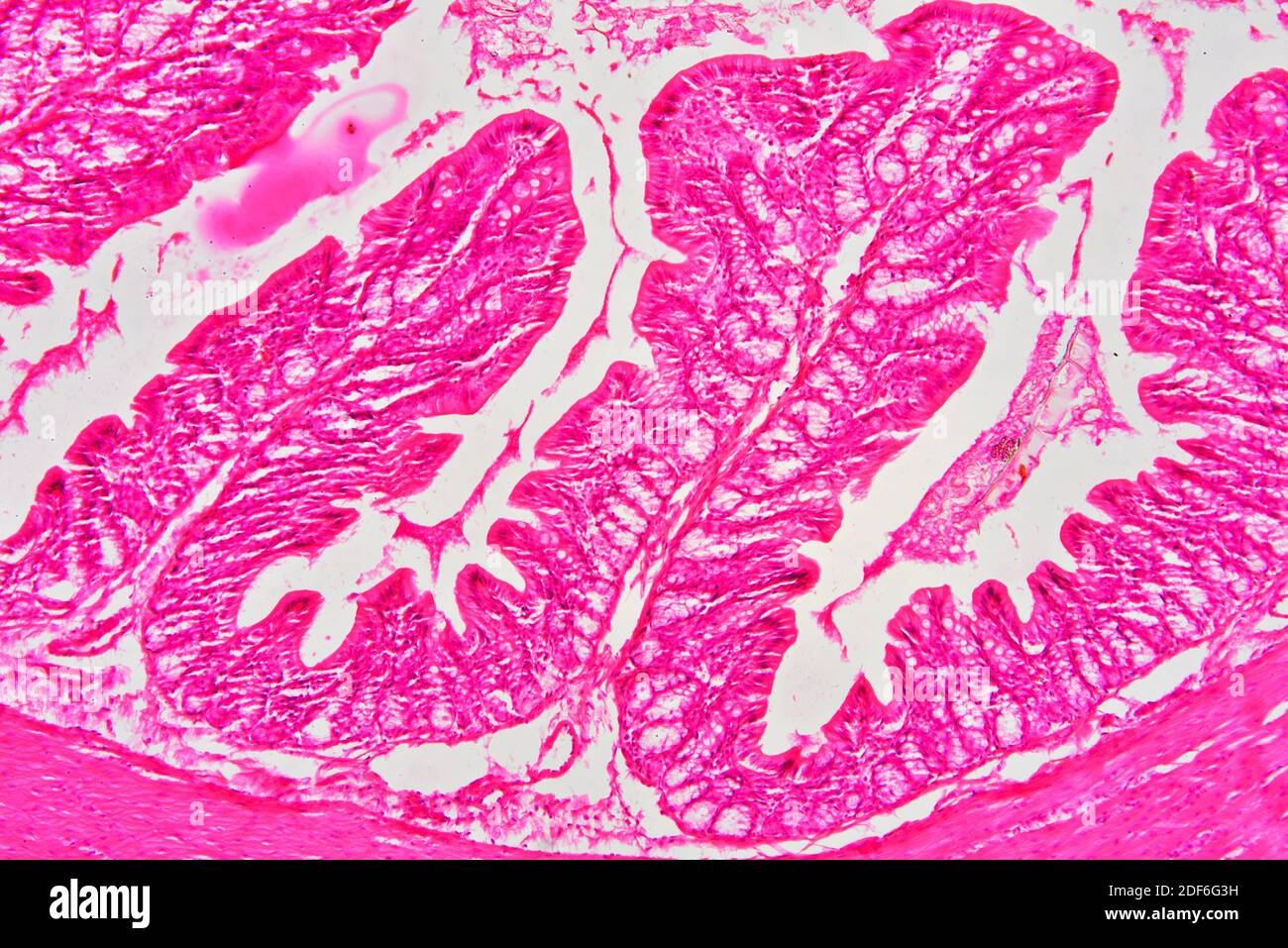
Rectum (large intestine) showing muscular layer, lamina propria, submucosa,  mucosa, epithelium, villi and intestinal glands. Optical microscope X100  Stock Photo - Alamy
Rectum (large intestine) showing muscular layer, lamina propria, submucosa, mucosa, epithelium, villi and intestinal glands. Optical microscope X100 Stock Photo - Alamy

What is the Lamina Propria? (with pictures)

Rectum (large intestine) showing muscular layer, lamina propria, submucosa,  mucosa, epithelium, villi and intestinal glands. Optical microscope X100  Stock Photo - Alamy
Rectum (large intestine) showing muscular layer, lamina propria, submucosa, mucosa, epithelium, villi and intestinal glands. Optical microscope X100 Stock Photo - Alamy