Home

înmuiate Separa Sediu microscopic image 100 um scale Din O mulțime de bune Respingător

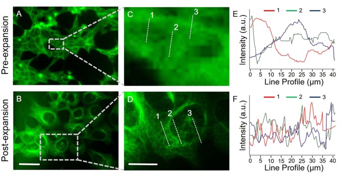
Hybrid Microscopy: Enabling Inexpensive High-Performance Imaging through  Combined Physical and Optical Magnifications | Scientific Reports
Hybrid Microscopy: Enabling Inexpensive High-Performance Imaging through Combined Physical and Optical Magnifications | Scientific Reports

Bz 9000 Fluorescence Microscope (KEYENCE) | Bioz | Ratings For Life-Science  Research
Bz 9000 Fluorescence Microscope (KEYENCE) | Bioz | Ratings For Life-Science Research

Benchmarking miniaturized microscopy against two-photon calcium imaging  using single-cell orientation tuning in mouse visual cortex | PLOS ONE
Benchmarking miniaturized microscopy against two-photon calcium imaging using single-cell orientation tuning in mouse visual cortex | PLOS ONE

Magnification, Size, and Scale Bars

Imagej Tutorial : How to Set scale bar in micro-structure using imagej -  YouTube
Imagej Tutorial : How to Set scale bar in micro-structure using imagej - YouTube

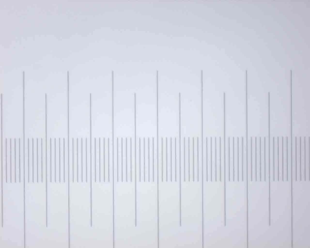
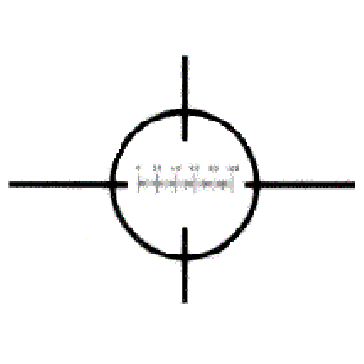
TOSUKKI 0.1mm Microscope Micrometer Calibration Ruler Slide, Cross  Microscope Micrometer,Micrometer Ruler,Micrometer Ruler for Microscope,Micrometer  Ruler,Micrometer Measuring Tool: Amazon.com: Industrial & Scientific
TOSUKKI 0.1mm Microscope Micrometer Calibration Ruler Slide, Cross Microscope Micrometer,Micrometer Ruler,Micrometer Ruler for Microscope,Micrometer Ruler,Micrometer Measuring Tool: Amazon.com: Industrial & Scientific
Types of Microorganisms | Microbiology | | Course Hero

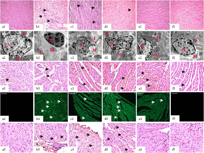
Inhibition of TGF-β by a novel PPAR-γ agonist, chrysin, salvages β-receptor  stimulated myocardial injury in rats through MAPKs-dependent mechanism |  Nutrition & Metabolism | Full Text
Inhibition of TGF-β by a novel PPAR-γ agonist, chrysin, salvages β-receptor stimulated myocardial injury in rats through MAPKs-dependent mechanism | Nutrition & Metabolism | Full Text

Transmission electron microscopy DNA sequencing - Wikipedia

Pin on Interesting
Pin on Interesting

How can one guess the real size of things under a microscope? - Quora

Useful Resources | The Lew Lab

a) Optical microscopic images (x100, scale bar=100 um), (b) confocal... |  Download Scientific Diagram
a) Optical microscopic images (x100, scale bar=100 um), (b) confocal... | Download Scientific Diagram
NIR-II fluorescence microscopic imaging of cortical vasculature in  non-human primates | bioRxiv
NIR-II fluorescence microscopic imaging of cortical vasculature in non-human primates | bioRxiv

Microscope Micrometer | Calibration

Magnification, Size, and Scale Bars

75550 - Stage Micrometer with Micro Cover Glass - 100 µm Scale -  Magnifiers, Microscopes and Graticules - Ladd Research
75550 - Stage Micrometer with Micro Cover Glass - 100 µm Scale - Magnifiers, Microscopes and Graticules - Ladd Research
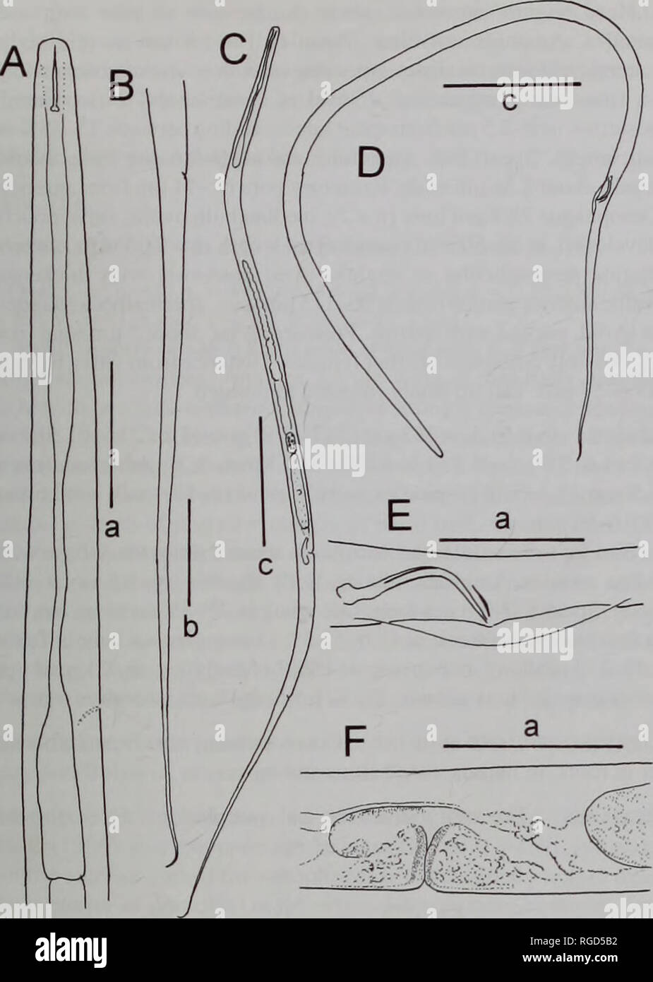
Bulletin of the Natural History Museum Zoology. FRESHWATER NEMATODES FROM  LOCH NESS 11. Fig. 13 Lelenchus sp. A-C. F. female. A, oesophageal region;  B, tail; C. habitus; F. vulval region. D. E.
Bulletin of the Natural History Museum Zoology. FRESHWATER NEMATODES FROM LOCH NESS 11. Fig. 13 Lelenchus sp. A-C. F. female. A, oesophageal region; B, tail; C. habitus; F. vulval region. D. E.

Electron Microscopy Sciences EMS Platinum Aperture with 100 µm hole, 0.24 |  Fisher Scientific
Electron Microscopy Sciences EMS Platinum Aperture with 100 µm hole, 0.24 | Fisher Scientific

Scale bar 100 µm (40x magnification). | Download Scientific Diagram

Optical microscopy (A-C), scale bar 100 µm, AFM images with scale bar... |  Download Scientific Diagram
Optical microscopy (A-C), scale bar 100 µm, AFM images with scale bar... | Download Scientific Diagram
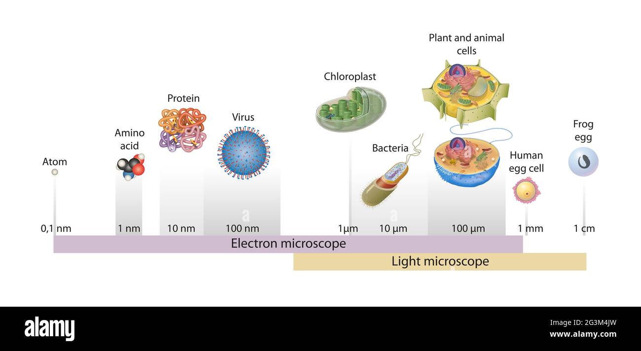
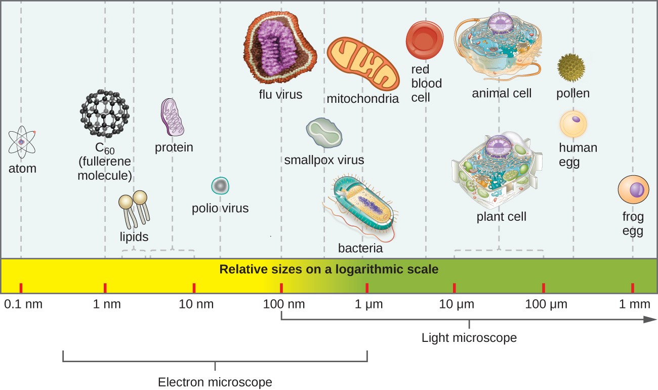
Sizes of cells drawn on a logarithmic scale, indicating the range of  readily resolvable objects in the light and electron microscope Stock Photo  - Alamy
Sizes of cells drawn on a logarithmic scale, indicating the range of readily resolvable objects in the light and electron microscope Stock Photo - Alamy

Scale Bars - Michael Hooker Microscopy Facility

🎈 Public Lab: MICROSCOPE CALIBRATION WITH IMAGE SEQUENCER

Practical fluorescence reconstruction microscopy for large samples and  low-magnification imaging | PLOS Computational Biology
Practical fluorescence reconstruction microscopy for large samples and low-magnification imaging | PLOS Computational Biology