Home

Din cand in cand Curând Evaluare mitochondria under light microscope spre exil usturoi grămadă

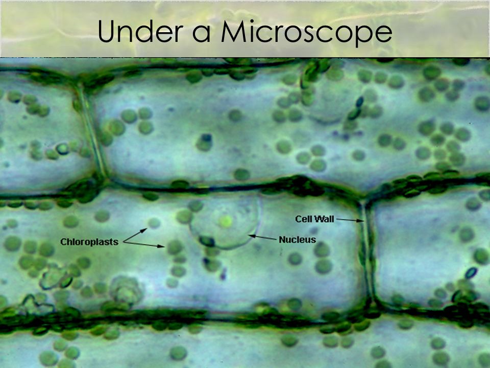
What Living Things You Can See Under a Light Microscope? - Rs' Science

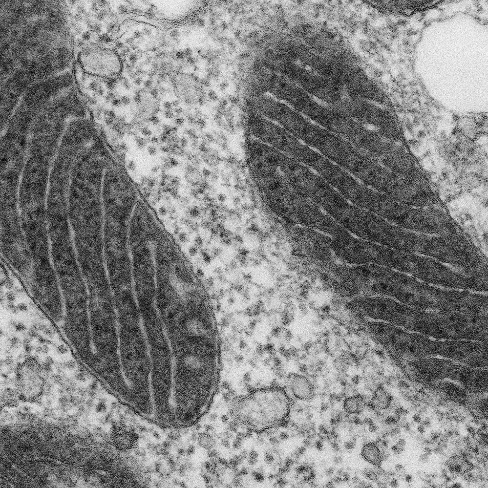
Mitochondria under the microscope — Science Learning Hub

Mitochondria live under Microscope - YouTube

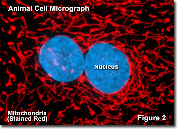
Molecular Expressions Cell Biology: Mitochondria

When we see a cheek cell through a microscope, can we see the mitochondria?  - Quora
When we see a cheek cell through a microscope, can we see the mitochondria? - Quora

Mitochondria Protection after Acute Ischemia Prevents Prolonged  Upregulation of IL-1β and IL-18 and Arrests CKD | American Society of  Nephrology
Mitochondria Protection after Acute Ischemia Prevents Prolonged Upregulation of IL-1β and IL-18 and Arrests CKD | American Society of Nephrology

The morphology of mitochondria. (a) Thin-section electron micrograph of...  | Download Scientific Diagram
The morphology of mitochondria. (a) Thin-section electron micrograph of... | Download Scientific Diagram

A new glow for electron microscopy | MIT News | Massachusetts Institute of  Technology
A new glow for electron microscopy | MIT News | Massachusetts Institute of Technology

Electron micrograph of a mitochondrion in a cell of the bat pancreas,... |  Download Scientific Diagram
Electron micrograph of a mitochondrion in a cell of the bat pancreas,... | Download Scientific Diagram

Live mitochondria seen in unprecedented detail: photobleaching in STED  microscopy overcome
Live mitochondria seen in unprecedented detail: photobleaching in STED microscopy overcome

Cell Biology for the Histologist | histology

Electron microscopy morphology of the mitochondrial network in gliomas and  their vascular microenvironment - ScienceDirect
Electron microscopy morphology of the mitochondrial network in gliomas and their vascular microenvironment - ScienceDirect

Light and electron microscopy showing ultrastructural changes in the... |  Download Scientific Diagram
Light and electron microscopy showing ultrastructural changes in the... | Download Scientific Diagram

Light and electron microscopy of mitochondria in the oocytes of Argulus...  | Download Scientific Diagram
Light and electron microscopy of mitochondria in the oocytes of Argulus... | Download Scientific Diagram

Mitochondria under the microscope | Mitochondria, Cell biology, Membrane  structure
Mitochondria under the microscope | Mitochondria, Cell biology, Membrane structure

Can you expect to see mitochondria while using a light compound microscope?  | Socratic
Can you expect to see mitochondria while using a light compound microscope? | Socratic

Mitochondria: A worthwhile object for ultrastructural qualitative  characterization and quantification of cells at physiological and  pathophysiological states using conventional transmission electron  microscopy - ScienceDirect
Mitochondria: A worthwhile object for ultrastructural qualitative characterization and quantification of cells at physiological and pathophysiological states using conventional transmission electron microscopy - ScienceDirect
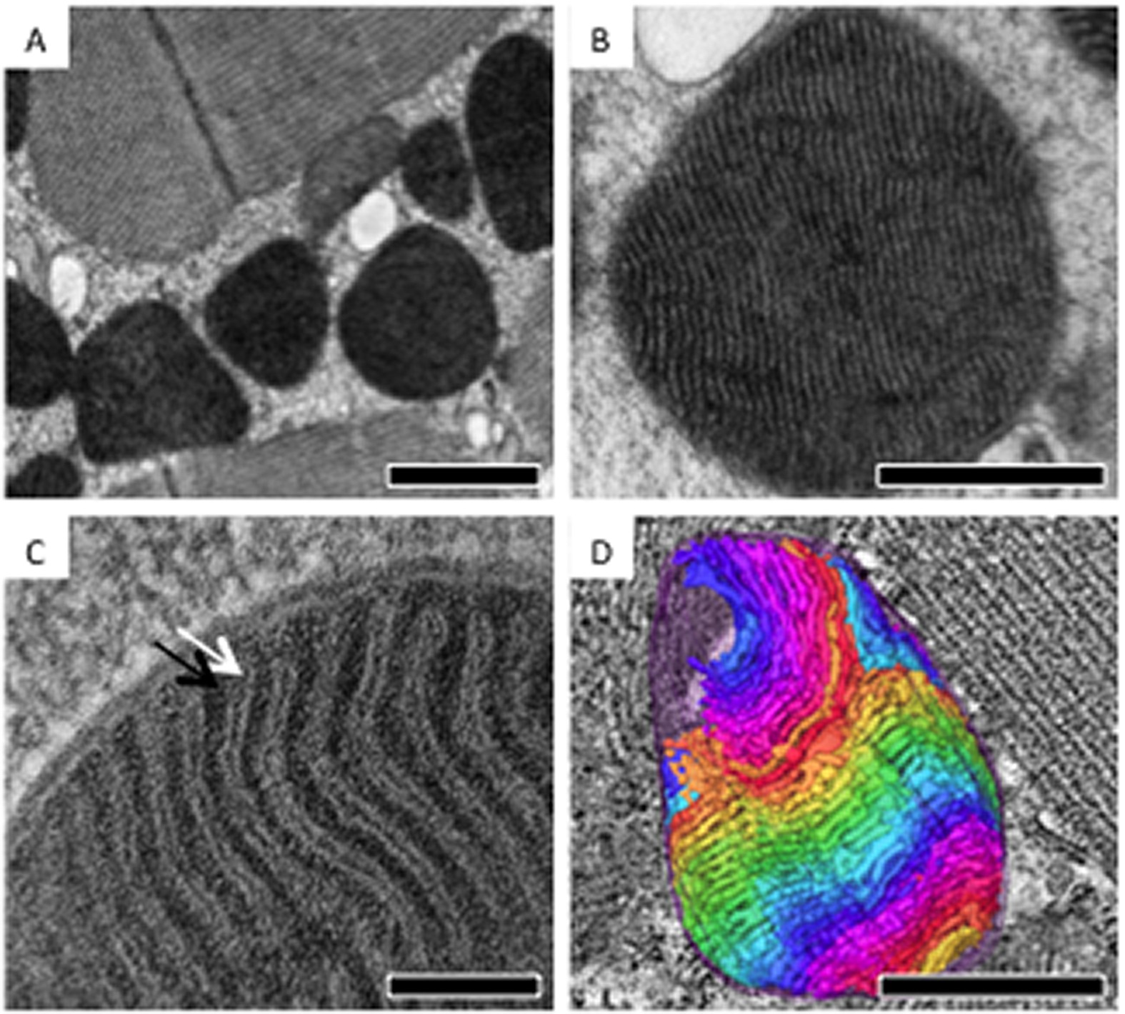
Mitochondria appearance under electron microscope (EM × 6000); A :... |  Download Scientific Diagram
Mitochondria appearance under electron microscope (EM × 6000); A :... | Download Scientific Diagram

Electron tomographic analysis reveals ultrastructural features of  mitochondrial cristae architecture which reflect energetic state and aging  | Scientific Reports
Electron tomographic analysis reveals ultrastructural features of mitochondrial cristae architecture which reflect energetic state and aging | Scientific Reports

What Living Things You Can See Under a Light Microscope? - Rs' Science

Mitochondria under the microscope — Science Learning Hub

Mitochondria: Power Producers in Cells

A LIGHT AND ELECTRON MICROSCOPE STUDY OF THE MORPHOLOGICAL CHANGES INDUCED  IN RAT LIVER CELLS BY THE AZO DYE 2-ME-DAB
A LIGHT AND ELECTRON MICROSCOPE STUDY OF THE MORPHOLOGICAL CHANGES INDUCED IN RAT LIVER CELLS BY THE AZO DYE 2-ME-DAB

Electron microscopy morphology of the mitochondrial network in gliomas and  their vascular microenvironment - ScienceDirect
Electron microscopy morphology of the mitochondrial network in gliomas and their vascular microenvironment - ScienceDirect

Mitochondrion, SEM - Stock Image - C001/2592 - Science Photo Library

Tem mitochondria hi-res stock photography and images - Alamy

Mitochondrion, TEM - Stock Image - C017/4146 - Science Photo Library