Home

indigen salariu Crustă phagocytosis microscope Fascinant Rafinat Nava spatiala

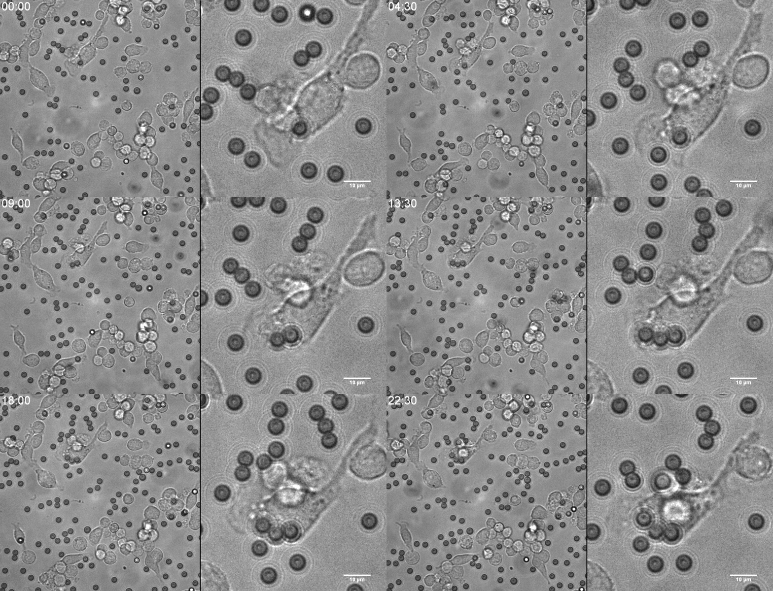
Light microscopy of in vivo phagocytosis from the peritoneal washes... |  Download Scientific Diagram
Light microscopy of in vivo phagocytosis from the peritoneal washes... | Download Scientific Diagram

Phagocytosis in a Shape-shifting Bacterium: Trends in Microbiology

Do all cells do phagocytosis? - Quora

Immune Cells Eating Bacteria (Phagocytosis) - YouTube

Phagocytosis Under the microscope - YouTube

phagocytosis with raw cells

Orientia tsutsugamushi Bacteria, Phagocytosis, TEM - Stock Image -  C043/4606 - Science Photo Library
Orientia tsutsugamushi Bacteria, Phagocytosis, TEM - Stock Image - C043/4606 - Science Photo Library

Electron microscopy images of monocyte-derived macrophages (MDM)... |  Download Scientific Diagram
Electron microscopy images of monocyte-derived macrophages (MDM)... | Download Scientific Diagram

Transmission electron microscopy. Phagocytosis of latex microspheres... |  Download Scientific Diagram
Transmission electron microscopy. Phagocytosis of latex microspheres... | Download Scientific Diagram

Phagocytosis
Phagocytosis

Phagocytes - Phagocytic cells - TeachMePhysiology

Premium Photo | Leukocyte phagocytosis, white blood cells in vein.  protection against viruses and bacteria. omicron strain. view of  neutrophils under the microscope. 3d render.
Premium Photo | Leukocyte phagocytosis, white blood cells in vein. protection against viruses and bacteria. omicron strain. view of neutrophils under the microscope. 3d render.

Figure 4 from Phagocytosis of bacteria by polymorphonuclear leukocytes: a  freeze-fracture, scanning electron microscope, and thin-section  investigation of membrane structure | Semantic Scholar
Figure 4 from Phagocytosis of bacteria by polymorphonuclear leukocytes: a freeze-fracture, scanning electron microscope, and thin-section investigation of membrane structure | Semantic Scholar

Microscopic materials hint at the origin of phagocytosis – Physics World

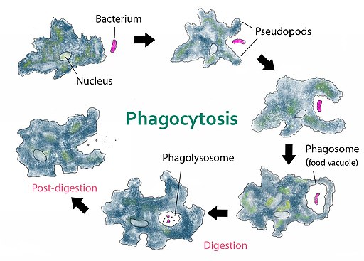
Phagocytosis - Definition, Steps, Process and Examples

Different Stages of Endocytosis (Phagocytosis) – Taking it INSIDE! |  Crawford Science & Technology Blog
Different Stages of Endocytosis (Phagocytosis) – Taking it INSIDE! | Crawford Science & Technology Blog

Platelet phagocytosis by neutrophils in a patient with antiphospholipid  syndrome | Rheumatology & Autoimmunity
Platelet phagocytosis by neutrophils in a patient with antiphospholipid syndrome | Rheumatology & Autoimmunity

Endocytosis Dr.Jastrow's electron microscopic atlas

Phagocytosis of Microorganisms

Transmission electron microscopy. Phagocytosis of latex microspheres... |  Download Scientific Diagram
Transmission electron microscopy. Phagocytosis of latex microspheres... | Download Scientific Diagram

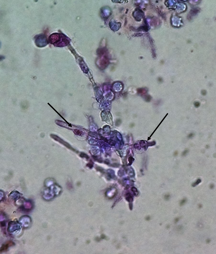
Urine Sediment of the Month: Identifying Phagocytosed Particles - Renal  Fellow Network
Urine Sediment of the Month: Identifying Phagocytosed Particles - Renal Fellow Network

Premium Photo | Leukocyte phagocytosis white blood cells in vein protection  against viruses and bacteria the concept of science and medicine view of  neutrophils under the microscope 3d render
Premium Photo | Leukocyte phagocytosis white blood cells in vein protection against viruses and bacteria the concept of science and medicine view of neutrophils under the microscope 3d render

phagocytosis | Definition, Process, & Examples | Britannica

PDF] Electron-microscope study of Dictyostelium discoideum plasma membrane  and its modifications during and after phagocytosis. | Semantic Scholar
PDF] Electron-microscope study of Dictyostelium discoideum plasma membrane and its modifications during and after phagocytosis. | Semantic Scholar
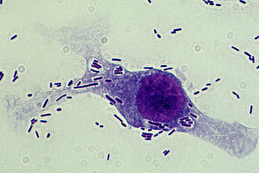
Tissue Macrophage Phagocytosis Photograph by Dennis Kunkel Microscopy/science  Photo Library - Fine Art America
Tissue Macrophage Phagocytosis Photograph by Dennis Kunkel Microscopy/science Photo Library - Fine Art America

Electron Microscopy
Electron Microscopy