Home

Girafă Permanent corect scanning electron microscope in romanian virtual Premedicație Monopol

Scanning Electron Microscope Technology from CamScan - Applied Beams LLC

Scanning Electron Microscope showing. (a) External view of female... |  Download Scientific Diagram
Scanning Electron Microscope showing. (a) External view of female... | Download Scientific Diagram

Electron Microscopy | Microscopy - U-M Biomedical Research Core Facilities

PDF) Potentialities of scanning electron microscopy and EDX analysis in  bullet wounds
PDF) Potentialities of scanning electron microscopy and EDX analysis in bullet wounds

Scanning electron microscope - Wikipedia

Benchtop and Compact Electron Microscopes

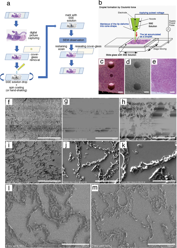
The NanoSuit method: a novel histological approach for examining paraffin  sections in a nondestructive manner by correlative light and electron  microscopy | Laboratory Investigation
The NanoSuit method: a novel histological approach for examining paraffin sections in a nondestructive manner by correlative light and electron microscopy | Laboratory Investigation

Scanning Electron Microscopy (SEM) Analysis and Imaging - TWI

Electron Microscopy | Microscopy - U-M Biomedical Research Core Facilities

Scanning electron microscope image detailing the biogenic (calcareous)... |  Download Scientific Diagram
Scanning electron microscope image detailing the biogenic (calcareous)... | Download Scientific Diagram

Scanning Electron Microscope Lets Students See World in “Different Light”

what does the scanning electron microscope use to magnify images

Core Imaging Instrumentation | Department of Biology | ECU

3D scanning electron microscopy for biology - Nanoanalysis - Oxford  Instruments
3D scanning electron microscopy for biology - Nanoanalysis - Oxford Instruments

Electron Microscopy | Microscopy - U-M Biomedical Research Core Facilities

Equipment
Equipment

In-Situ Workflow Provides Deeper Insights Into Material Properties For  Field Emission Scanning Electron Microscopes – Metrology and Quality News -  Online Magazine
In-Situ Workflow Provides Deeper Insights Into Material Properties For Field Emission Scanning Electron Microscopes – Metrology and Quality News - Online Magazine
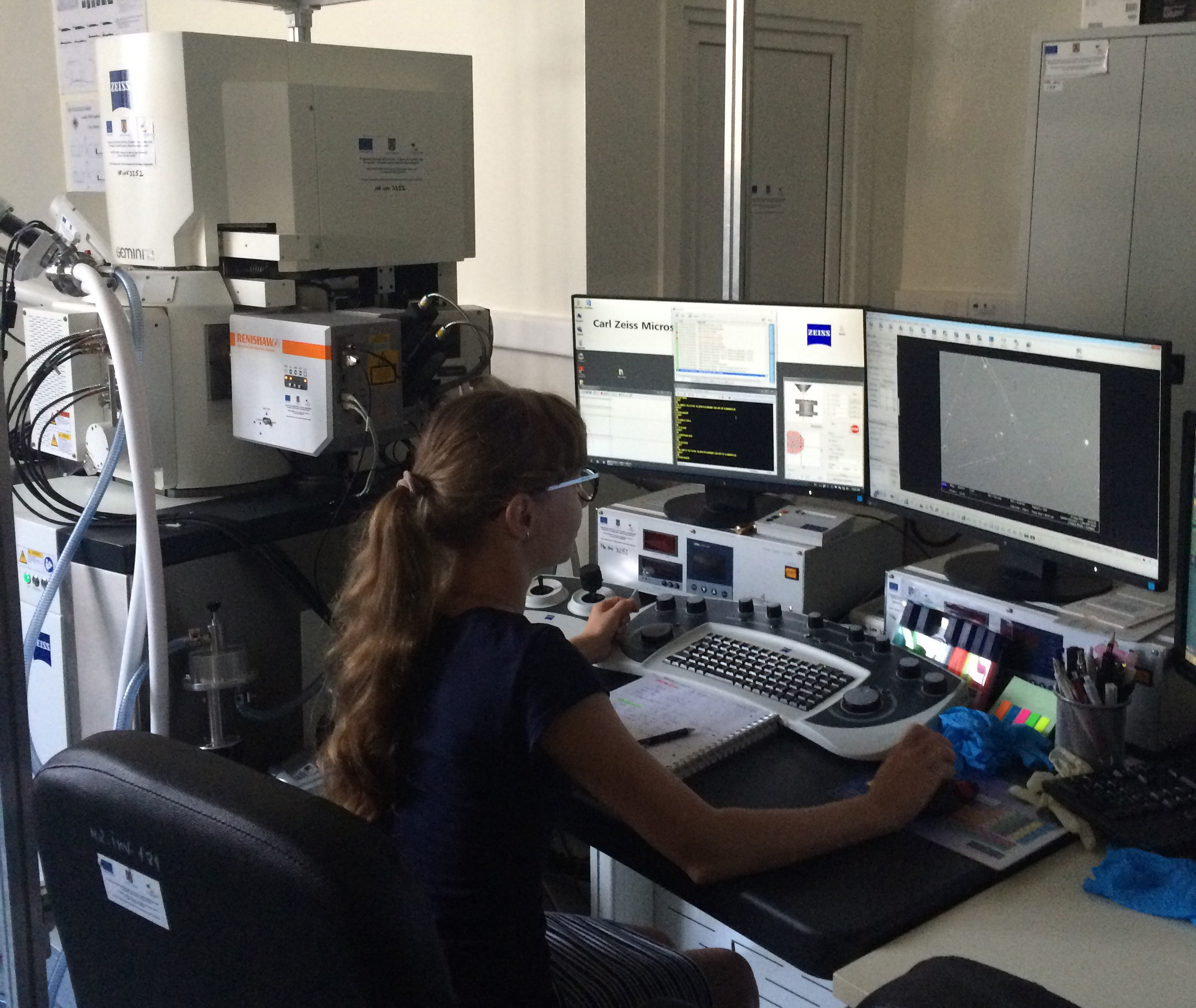
Combining Raman microscopy with scanning electron microscopy (SEM) to study  inorganic and mineral samples at the Geological Institute of Romania,  Bucharest
Combining Raman microscopy with scanning electron microscopy (SEM) to study inorganic and mineral samples at the Geological Institute of Romania, Bucharest

Scanning electron microscopy with 4000×. a Mordanted sample M14 with 5%...  | Download Scientific Diagram
Scanning electron microscopy with 4000×. a Mordanted sample M14 with 5%... | Download Scientific Diagram

Covalent Metrology Expands Scanning Electron Microscopy Capabilities –  Metrology and Quality News - Online Magazine
Covalent Metrology Expands Scanning Electron Microscopy Capabilities – Metrology and Quality News - Online Magazine
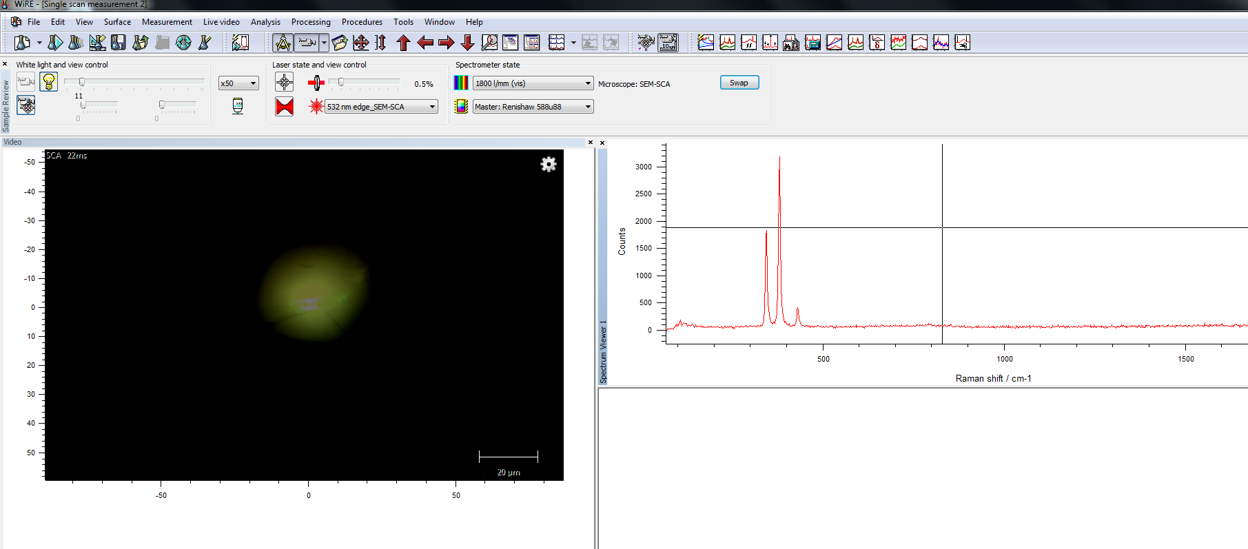
Combining Raman microscopy with scanning electron microscopy (SEM) to study  inorganic and mineral samples at the Geological Institute of Romania,  Bucharest
Combining Raman microscopy with scanning electron microscopy (SEM) to study inorganic and mineral samples at the Geological Institute of Romania, Bucharest

Electron Microscopy | Microscopy - U-M Biomedical Research Core Facilities