Home

perdea Groenlanda Metaforă skin surface microscope Arhiva Liniște Pocăinţă

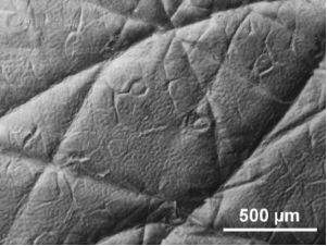
BJNANO - Surface topography and contact mechanics of dry and wet human skin

Premium Vector | Close up illustration of bacteria on surface of skin  mucous membrane or intestine under microscope
Premium Vector | Close up illustration of bacteria on surface of skin mucous membrane or intestine under microscope

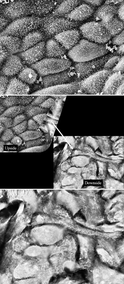
Applications of Electron Microscopy in Medicine - 2008 - Wiley Analytical  Science
Applications of Electron Microscopy in Medicine - 2008 - Wiley Analytical Science

SciencePhotoLibrary sur Twitter : "Your skin under a microscope! The top  layer is the stratum corneum (flaky, pale brown), dead skin cells that form  the surface of the skin. C:Eye of Science/SPL
SciencePhotoLibrary sur Twitter : "Your skin under a microscope! The top layer is the stratum corneum (flaky, pale brown), dead skin cells that form the surface of the skin. C:Eye of Science/SPL

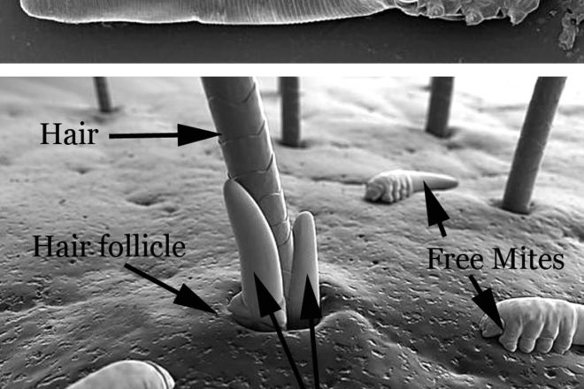
Surface of human skin with a hair follicle and squamous epithelium surface  ce… | Microscopic photography, Scanning electron microscope, Scanning  electron microscopy
Surface of human skin with a hair follicle and squamous epithelium surface ce… | Microscopic photography, Scanning electron microscope, Scanning electron microscopy
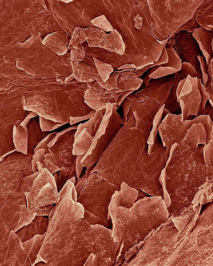
Amazing 27 Things Under The Microscope With Diagrams

Human Skin Epidermis Photograph by Dennis Kunkel Microscopy/science Photo  Library - Fine Art America
Human Skin Epidermis Photograph by Dennis Kunkel Microscopy/science Photo Library - Fine Art America
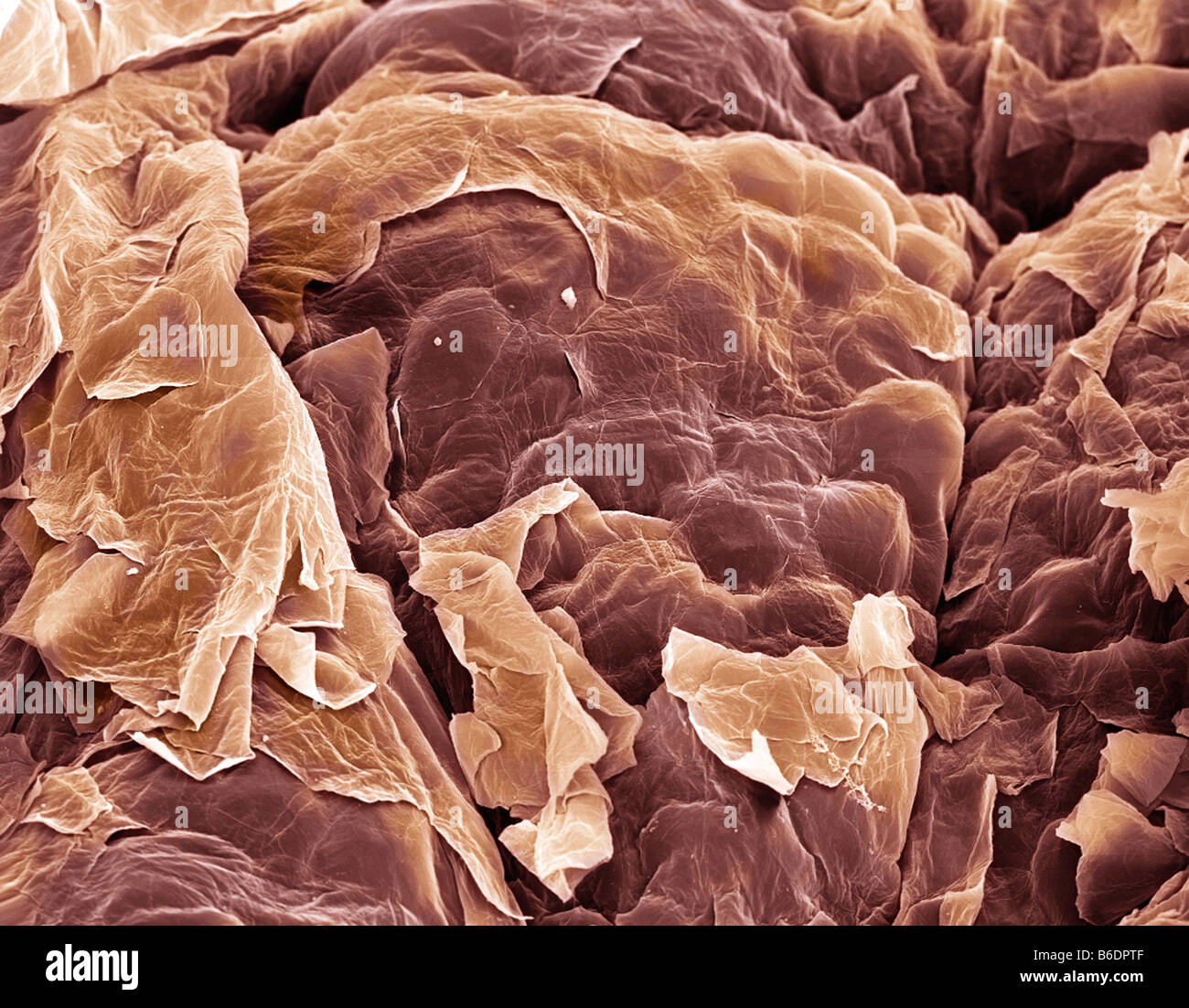
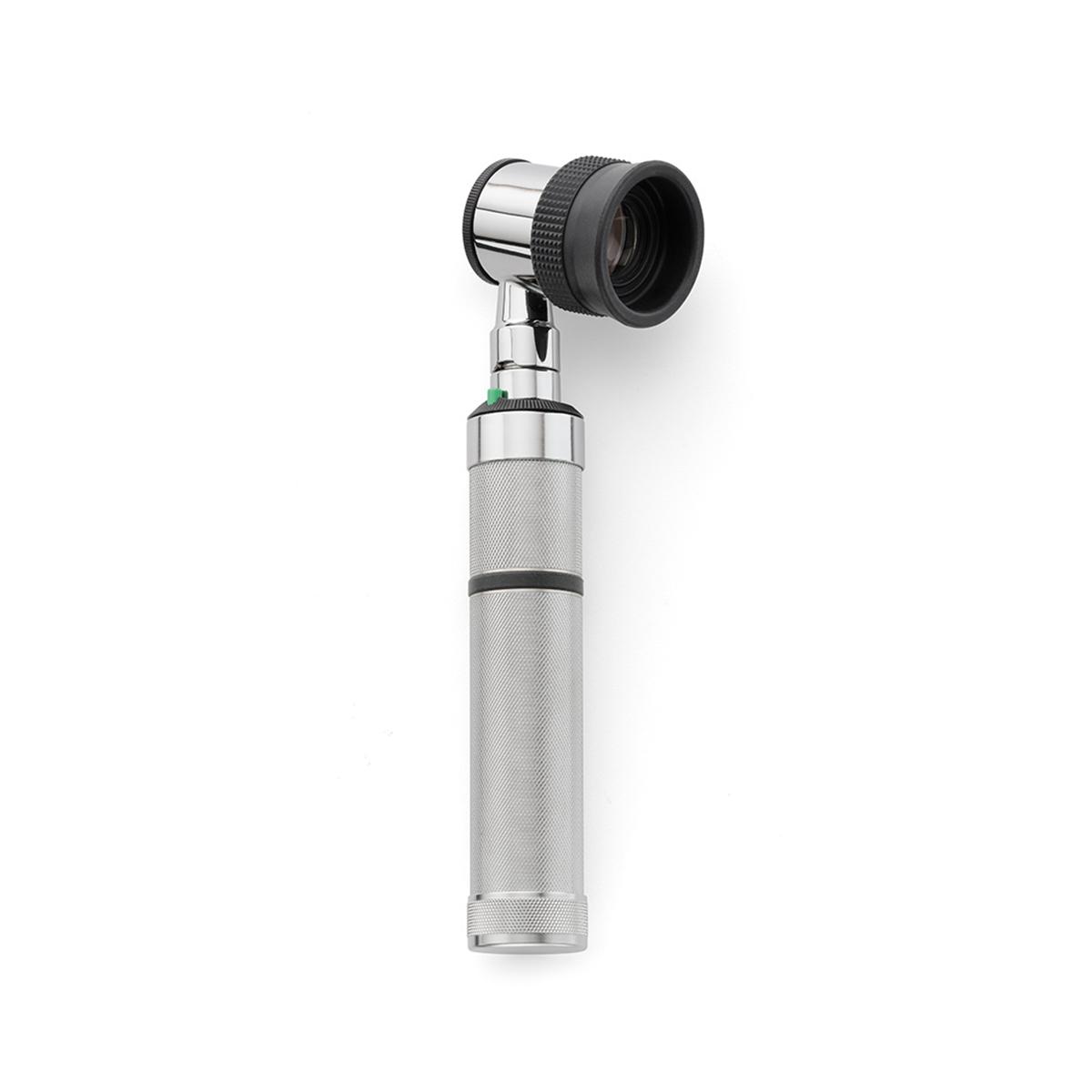
EpiScope Skin Surface Microscope | Welch Allyn | Hillrom

Incredible Inner Space: gallery

Scanning electron microscopy of the normal (left) and scar (right)... |  Download Scientific Diagram
Scanning electron microscopy of the normal (left) and scar (right)... | Download Scientific Diagram

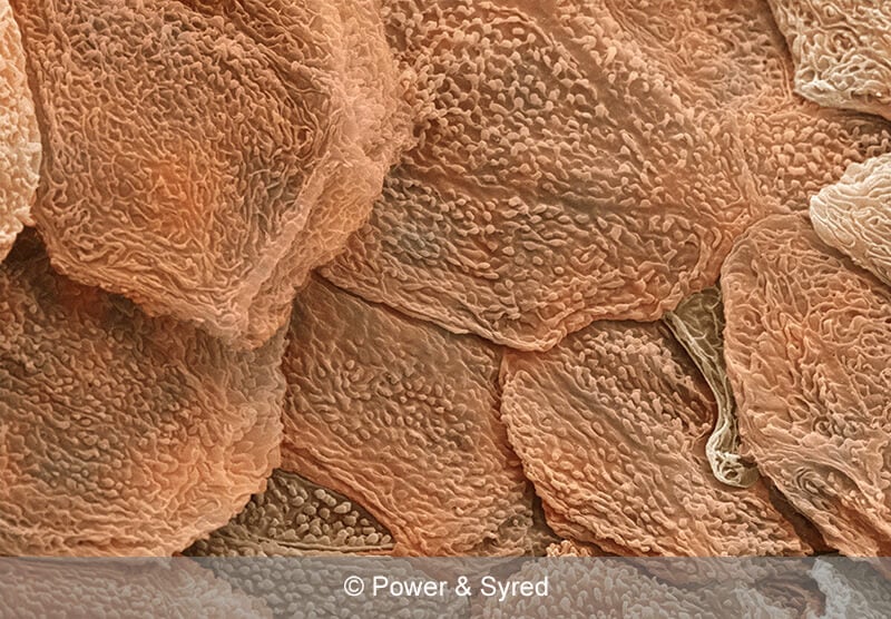
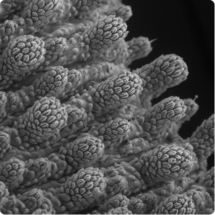
Skin. Coloured scanning electron micrograph (SEM) of squamous epithelial  cells on the skin surface Stock Photo - Alamy
Skin. Coloured scanning electron micrograph (SEM) of squamous epithelial cells on the skin surface Stock Photo - Alamy

Figure 4 from The stratum corneum barrier: the final frontier. | Semantic  Scholar
Figure 4 from The stratum corneum barrier: the final frontier. | Semantic Scholar

5,238 Skin Microscope Stock Photos, Pictures & Royalty-Free Images - iStock

Zoology with FurU - Surface of our skin😮 Microscopic view🔬 | Facebook

Snakeskin on a Microscopic Scale—Presenting the IOTY 2020 EMEA Regional  Winner | Olympus LS
Snakeskin on a Microscopic Scale—Presenting the IOTY 2020 EMEA Regional Winner | Olympus LS

Science Snippet: Skin Mites - Door County Pulse

Streptococci. Spherical bacteria on the surface of skin or mucous membrane,  model of staphylococcus and streptococcus, model of microbes, bacteria  simulating electron microscope, pyogenic bacteria Stock Illustration |  Adobe Stock
Streptococci. Spherical bacteria on the surface of skin or mucous membrane, model of staphylococcus and streptococcus, model of microbes, bacteria simulating electron microscope, pyogenic bacteria Stock Illustration | Adobe Stock

Bacteria Staphylococcus Aureus On The Surface Of Skin Or Mucous Membrane,  Model Of Staphylococcus, Superbug, MRSA, Model Of Microbes, Bacteria  Simulating Electron Microscope, Pyogenic Bacteria Stock Photo, Picture And  Royalty Free Image.
Bacteria Staphylococcus Aureus On The Surface Of Skin Or Mucous Membrane, Model Of Staphylococcus, Superbug, MRSA, Model Of Microbes, Bacteria Simulating Electron Microscope, Pyogenic Bacteria Stock Photo, Picture And Royalty Free Image.

sequential.skin - A section of Human skin layers @sciencephotolibrary  ⠀⠀⠀⠀⠀⠀⠀⠀⠀⠀⠀⠀ ⠀⠀⠀⠀⠀⠀⠀⠀⠀⠀⠀⠀ ⠀⠀⠀⠀⠀⠀⠀⠀⠀⠀⠀⠀ Coloured scanning electron  micrograph (SEM) of a section through human skin with a hair (upper left ...
sequential.skin - A section of Human skin layers @sciencephotolibrary ⠀⠀⠀⠀⠀⠀⠀⠀⠀⠀⠀⠀ ⠀⠀⠀⠀⠀⠀⠀⠀⠀⠀⠀⠀ ⠀⠀⠀⠀⠀⠀⠀⠀⠀⠀⠀⠀ Coloured scanning electron micrograph (SEM) of a section through human skin with a hair (upper left ...

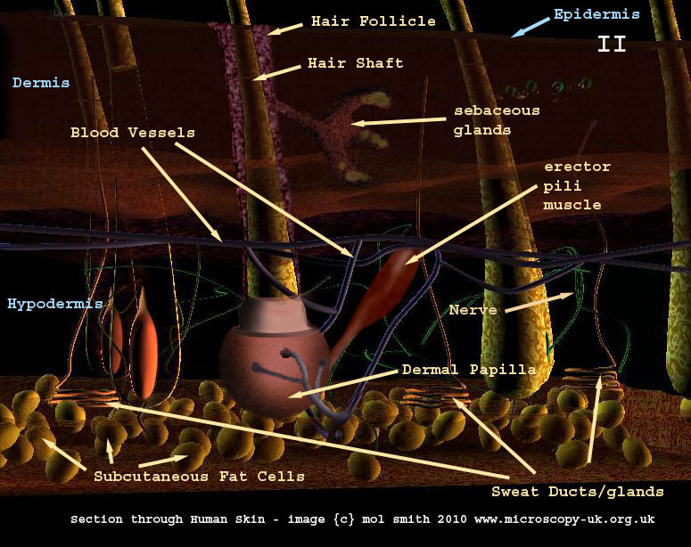
Human Cells - Part II an overview for light microscopists - Skin

Hair, Fungus, Skin Under Microscope Stock Illustration - Illustration of  loopable, generated: 157826737
Hair, Fungus, Skin Under Microscope Stock Illustration - Illustration of loopable, generated: 157826737
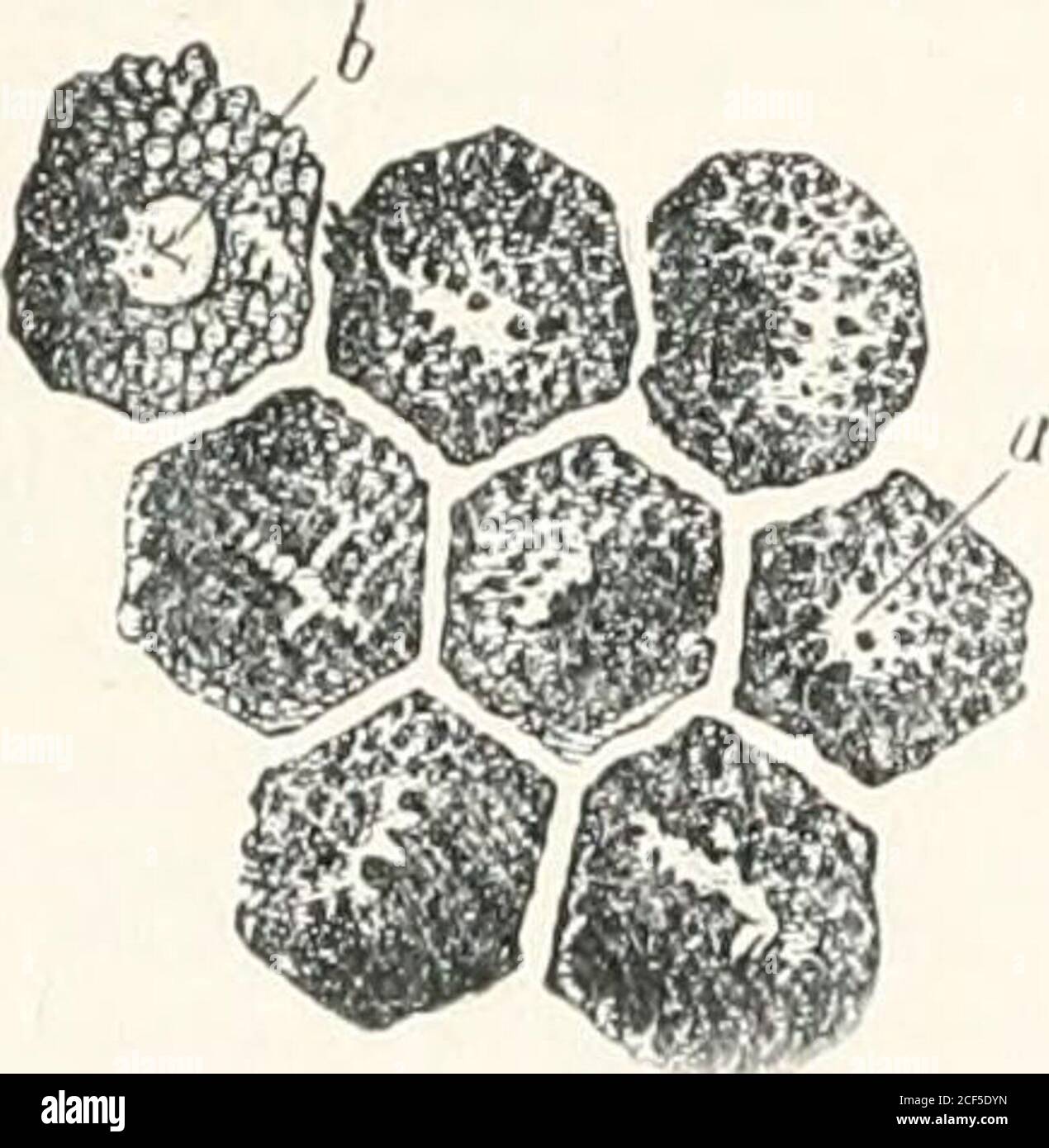
The microscope and its revelations. o be the peculiar seat of thecolour of  the skin ; it received the desig-nation of Malpighian layer or rete iniwosn  m.FIG. 776.—Cells from the pig-
The microscope and its revelations. o be the peculiar seat of thecolour of the skin ; it received the desig-nation of Malpighian layer or rete iniwosn m.FIG. 776.—Cells from the pig-

Scanning electron microscopy of the wound surface. (a) Intact skin,... |  Download Scientific Diagram
Scanning electron microscopy of the wound surface. (a) Intact skin,... | Download Scientific Diagram

PDF] The collagenic structure of human digital skin seen by scanning  electron microscopy after Ohtani maceration technique. | Semantic Scholar
PDF] The collagenic structure of human digital skin seen by scanning electron microscopy after Ohtani maceration technique. | Semantic Scholar