Home

stres dividend Nominal staphylococcus aureus microscope capodopera Decizie Spori

728 Staphylococcus Aureus Photos and Premium High Res Pictures - Getty  Images
728 Staphylococcus Aureus Photos and Premium High Res Pictures - Getty Images

Are there visual differences between the cells of methicillin-resistant staphylococcus  aureus (MRSA) and methicillin-susceptible S. aureus? I'm talking cell size,  opacity, coloration etc. Not colony characteristics. - Quora
Are there visual differences between the cells of methicillin-resistant staphylococcus aureus (MRSA) and methicillin-susceptible S. aureus? I'm talking cell size, opacity, coloration etc. Not colony characteristics. - Quora

New Understanding of 'Superantigens' Could Lead To Improved Staph Infection  Treatments - University of Wisconsin School of Veterinary Medicine
New Understanding of 'Superantigens' Could Lead To Improved Staph Infection Treatments - University of Wisconsin School of Veterinary Medicine

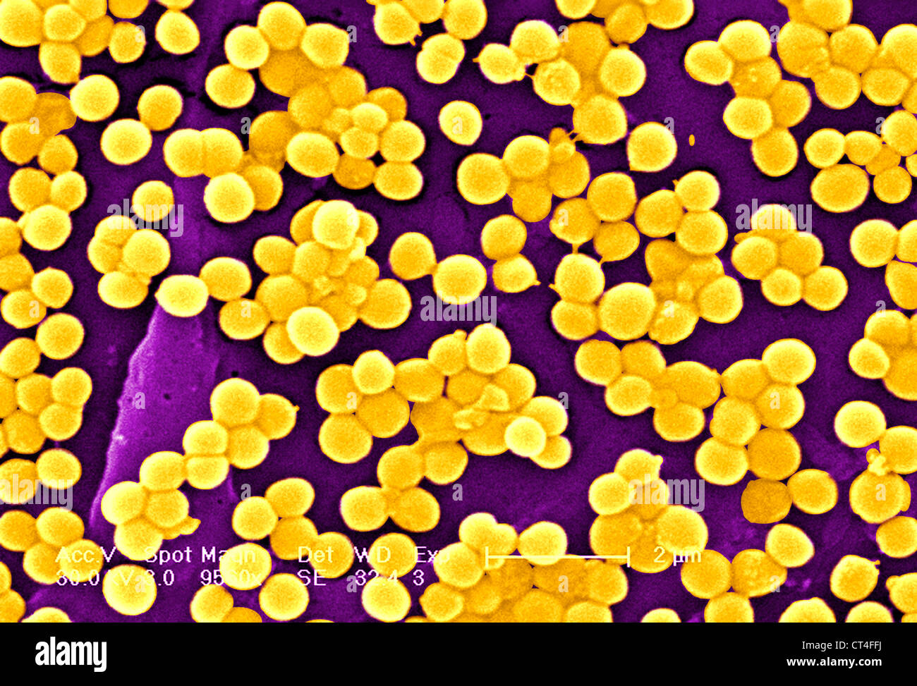
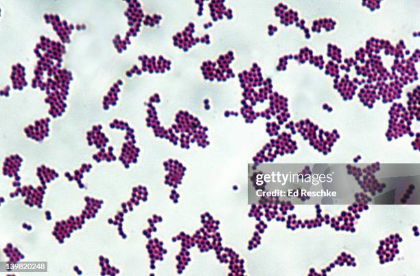
Staphylococcus hi-res stock photography and images - Alamy

Staphylococcus aureus light microscopy. Morphology of Staphylococcus aureus  under the microscope. Micrograph of S.aureus, Gram stain. Gram-stained  smear from culture.
Staphylococcus aureus light microscopy. Morphology of Staphylococcus aureus under the microscope. Micrograph of S.aureus, Gram stain. Gram-stained smear from culture.
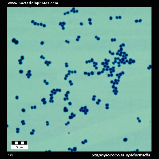
Staphylococcus epidermidis under microscope: microscopy of Gram-positive  cocci, morphology and microscopic appearance of Staphylococcus epidermidis,  S.epidermidis gram stain and colony morphology on agar, clinical  significance
Staphylococcus epidermidis under microscope: microscopy of Gram-positive cocci, morphology and microscopic appearance of Staphylococcus epidermidis, S.epidermidis gram stain and colony morphology on agar, clinical significance

Staphylococcus aureus under microscope: microscopy of Gram-positive cocci,  morphology and microscopic appearance of Staphylococcus aureus, S.aureus  gram stain and colony morphology on agar, clinical significance
Staphylococcus aureus under microscope: microscopy of Gram-positive cocci, morphology and microscopic appearance of Staphylococcus aureus, S.aureus gram stain and colony morphology on agar, clinical significance

Staphylococcus aureus | Gram positive stain under oil immers… | Flickr

Gram's staining of S. aureus (100X). Grapes like (black arrow) Gram... |  Download Scientific Diagram
Gram's staining of S. aureus (100X). Grapes like (black arrow) Gram... | Download Scientific Diagram

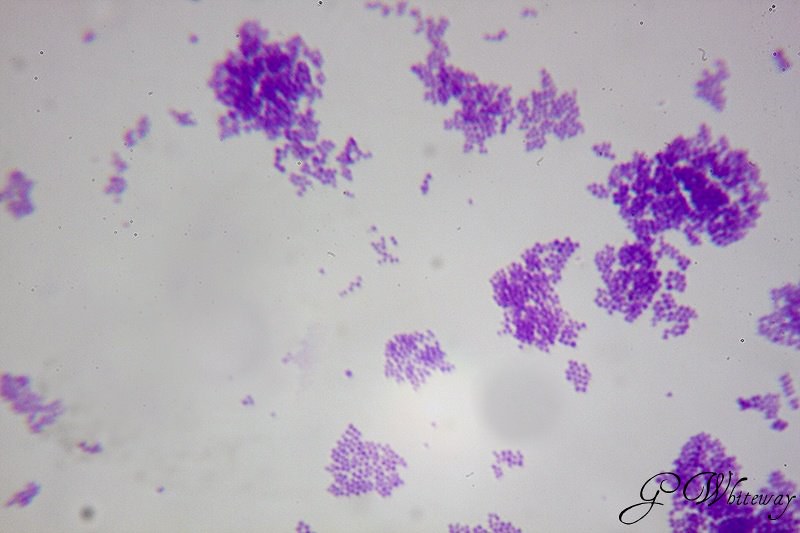
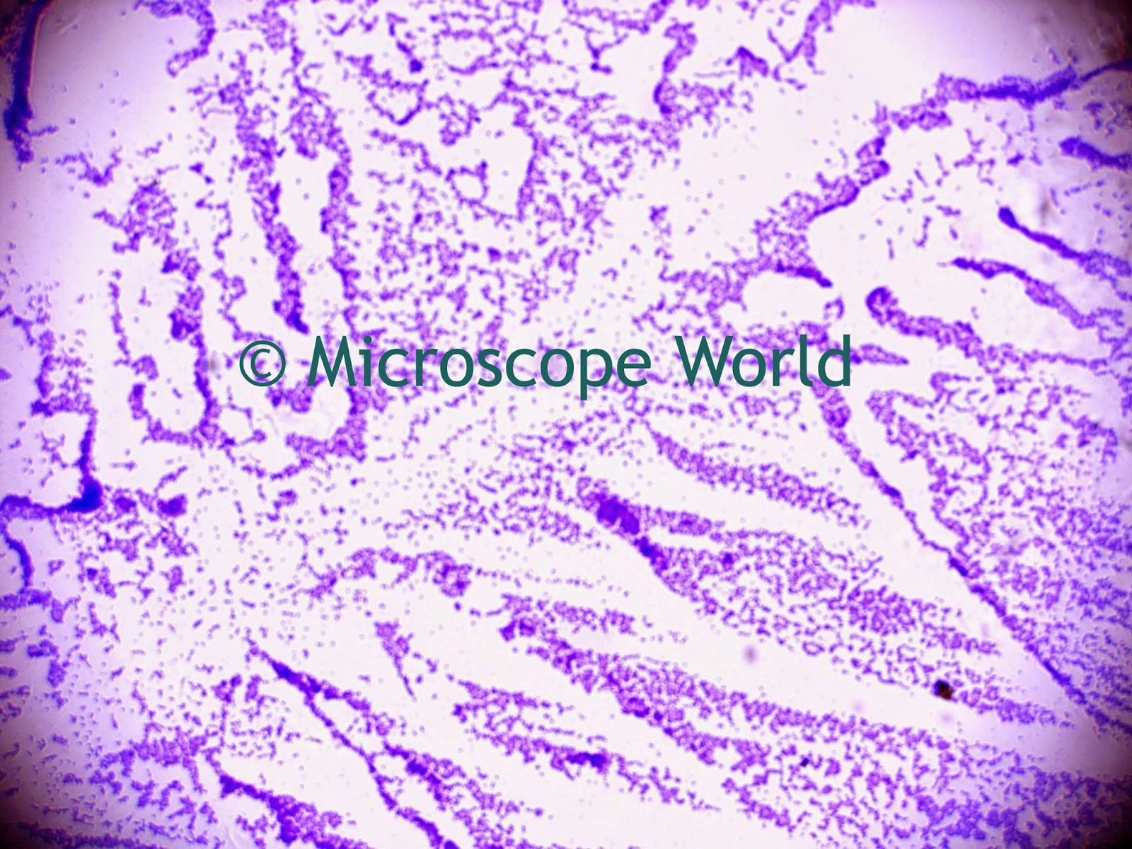
Microscope World Blog: Staphylococcus Under the Microscope

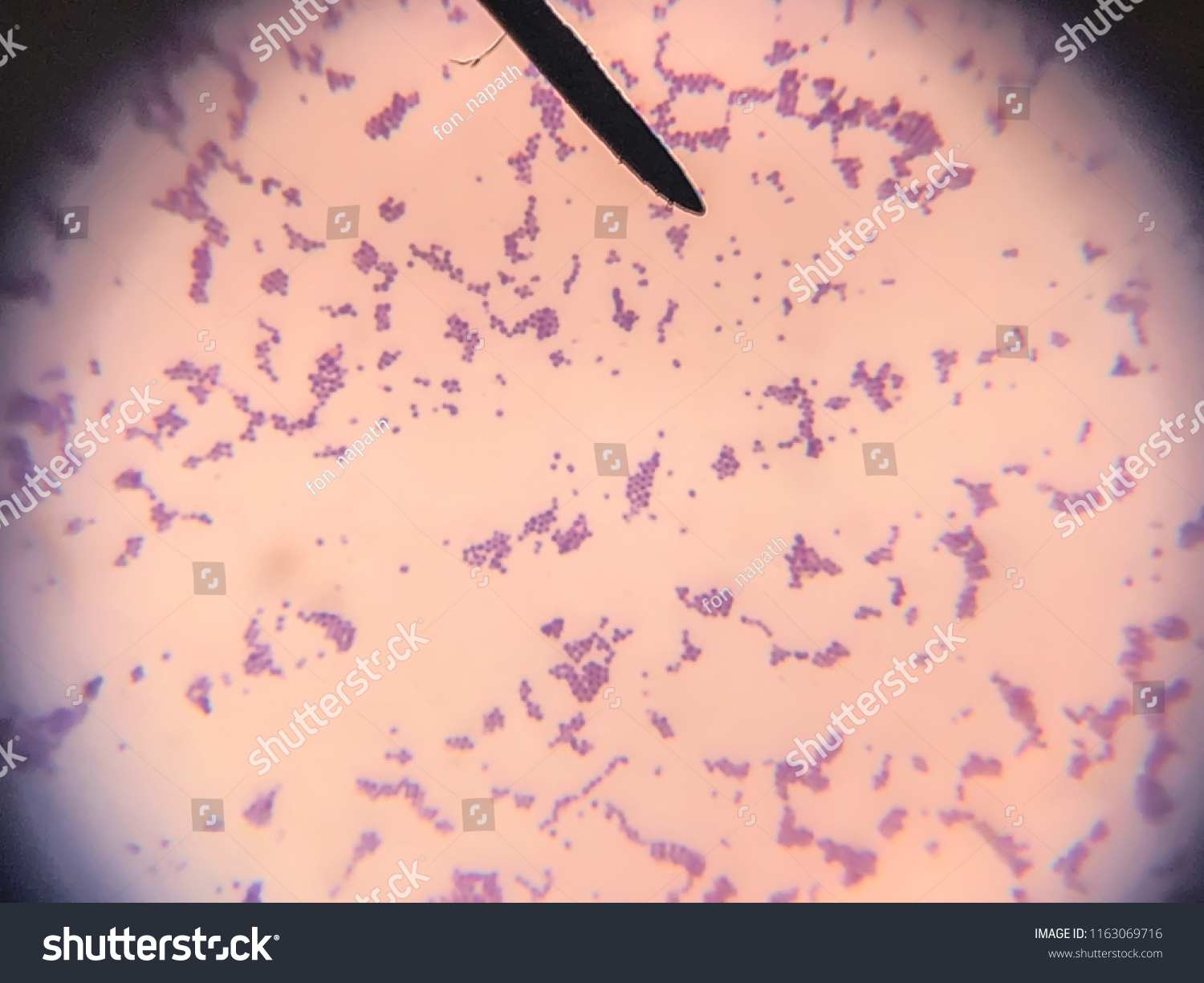
Morphology Bacteria Staphylococcus Aureus Cell Gram Stock Photo 1163069716  | Shutterstock
Morphology Bacteria Staphylococcus Aureus Cell Gram Stock Photo 1163069716 | Shutterstock

Microscope World Blog: Staphylococcus Under the Microscope

Details - Public Health Image Library(PHIL)

Eisco Prepared Microscope Slide - Staphylococcus Aureus Gram Positive  Microbiology | Fisher Scientific
Eisco Prepared Microscope Slide - Staphylococcus Aureus Gram Positive Microbiology | Fisher Scientific
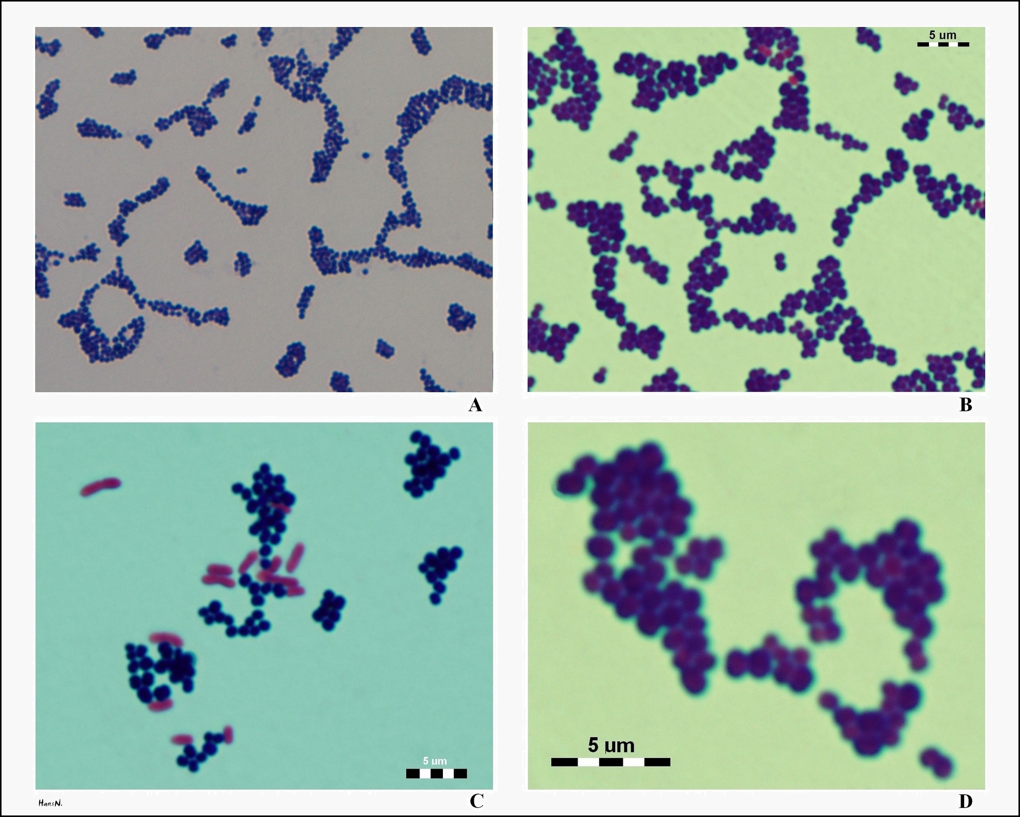
S. aureus under the microscope. Microscopic appearance and morphology of S. aureus. Cell arrangement.

Staphylococcus - Wikipedia

Staphylococcus aureus and Ecoli under microscope: microscopy of  Gram-positive cocci and Gram-negative bacilli, morphology and microscopic  appearance of Staphylococcus aureus and E.coli, S.aureus gram stain and  colony morphology on agar, clinical ...
Staphylococcus aureus and Ecoli under microscope: microscopy of Gram-positive cocci and Gram-negative bacilli, morphology and microscopic appearance of Staphylococcus aureus and E.coli, S.aureus gram stain and colony morphology on agar, clinical ...

Microbiology 201l > Hubert > Flashcards > Final Lab | StudyBlue |  Microbiology lab, Microbiology, Medical laboratory scientist
Microbiology 201l > Hubert > Flashcards > Final Lab | StudyBlue | Microbiology lab, Microbiology, Medical laboratory scientist

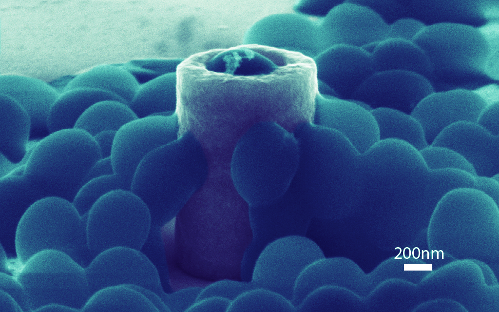
Scientists show which surfaces attract clingy Staph bacteria | Research UC  Berkeley
Scientists show which surfaces attract clingy Staph bacteria | Research UC Berkeley

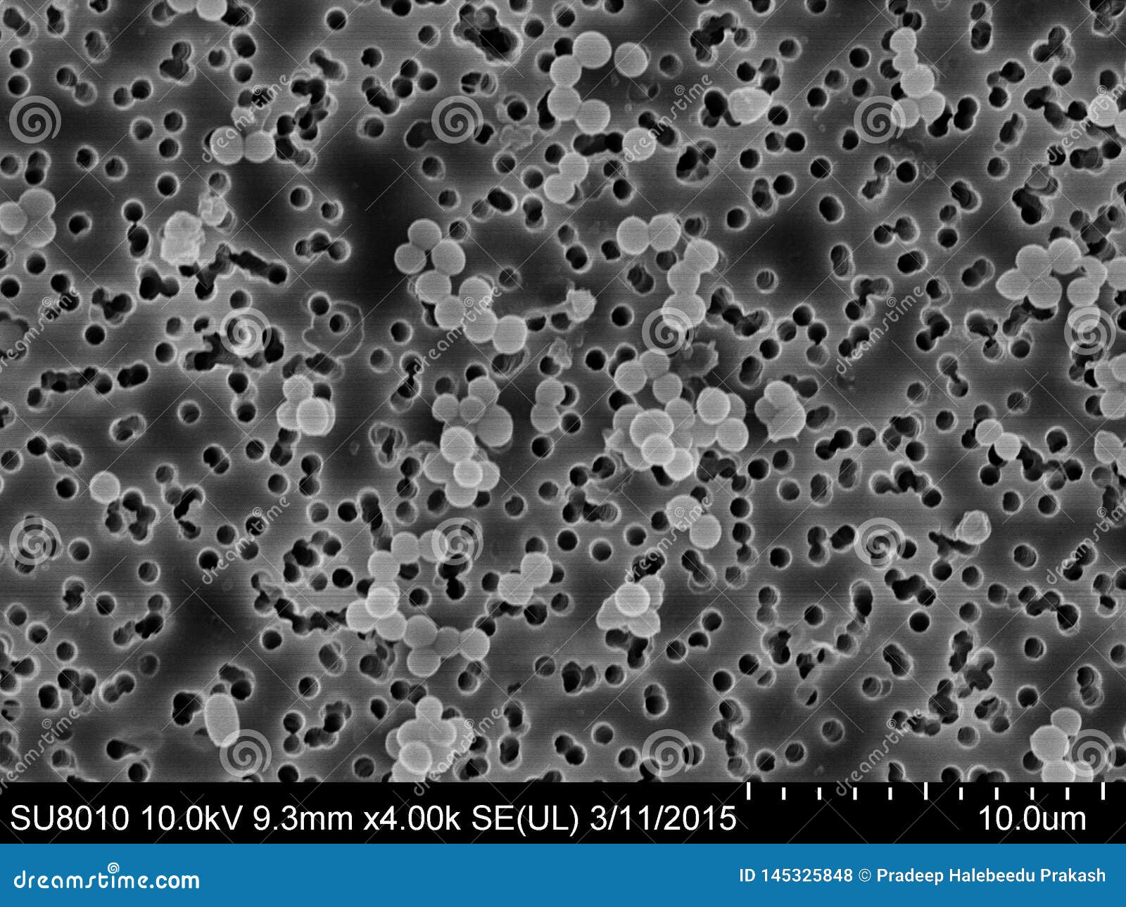
Staphylococcus Aureus on a Membrane Stock Photo - Image of hospital,  bacteria: 145325848
Staphylococcus Aureus on a Membrane Stock Photo - Image of hospital, bacteria: 145325848

Picture showing Staphylococcus aureus under the microscope | Download  Scientific Diagram
Picture showing Staphylococcus aureus under the microscope | Download Scientific Diagram

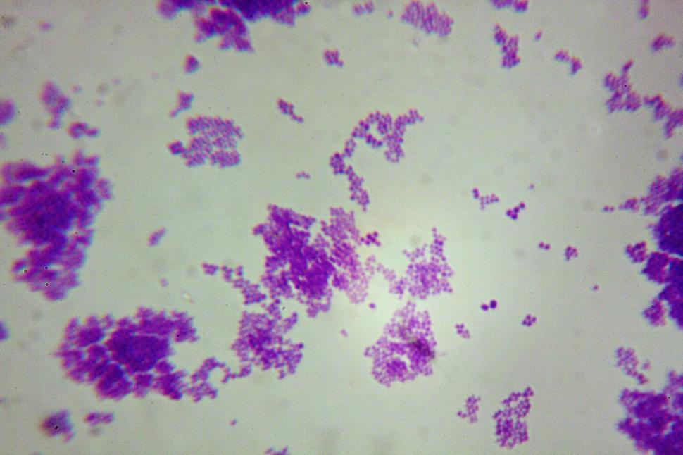
Free Stock Photo of Staphylococcus aureus | Download Free Images and Free  Illustrations
Free Stock Photo of Staphylococcus aureus | Download Free Images and Free Illustrations

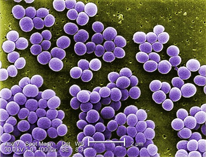
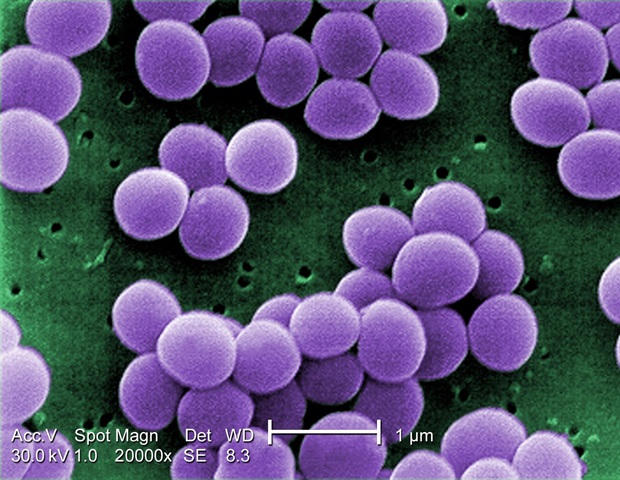
Staphylococcus aureus Bacteria | Scanning electron micrograp… | Flickr

Staphylococcus Aureus Microbiology

Staphylococcus aureus Slide, w.m.: Science Lab Microbiology Supplies:  Amazon.com: Industrial & Scientific
Staphylococcus aureus Slide, w.m.: Science Lab Microbiology Supplies: Amazon.com: Industrial & Scientific

Staphylococcus epidermidis 400x | Marc Perkins Photography