Home

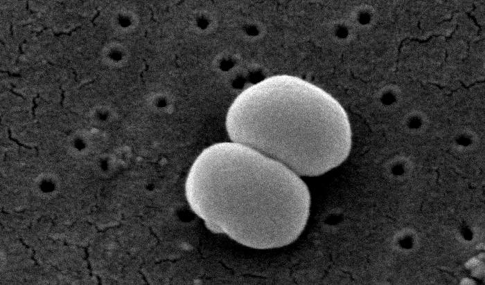
Pana cand aproape sticla staphylococcus epidermidis under microscope Special cu laser Pana acum

Staphylococcus aureus seen under microscope after Gram's staining |  Download Scientific Diagram
Staphylococcus aureus seen under microscope after Gram's staining | Download Scientific Diagram

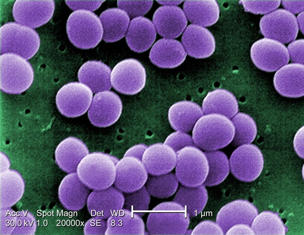
Staphylococcus epidermidis | bacterium | Britannica

Effect of polyherbal microemulsion on Staphylococcus epidermidis:  Formulation development, CCD based optimization, characterization, and  antibacterial activity by scanning electron microscopy - ScienceDirect
Effect of polyherbal microemulsion on Staphylococcus epidermidis: Formulation development, CCD based optimization, characterization, and antibacterial activity by scanning electron microscopy - ScienceDirect

Gram's staining of S. aureus (100X). Grapes like (black arrow) Gram... |  Download Scientific Diagram
Gram's staining of S. aureus (100X). Grapes like (black arrow) Gram... | Download Scientific Diagram

Staphylococcus aureus and Ecoli under microscope: microscopy of  Gram-positive cocci and Gram-negative bacilli, morphology and microscopic  appearance of Staphylococcus aureus and E.coli, S.aureus gram stain and  colony morphology on agar, clinical ...
Staphylococcus aureus and Ecoli under microscope: microscopy of Gram-positive cocci and Gram-negative bacilli, morphology and microscopic appearance of Staphylococcus aureus and E.coli, S.aureus gram stain and colony morphology on agar, clinical ...

Staphylococcus - Wikipedia

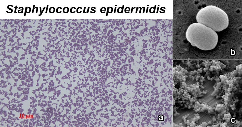
Staphylococcus epidermidis- An Overview - Microbe Notes

Staphylococcus aureus and Staphylococcus epidermidis Virulence Strains as  Causative Agents of Persistent Infections in Breast Implants | PLOS ONE
Staphylococcus aureus and Staphylococcus epidermidis Virulence Strains as Causative Agents of Persistent Infections in Breast Implants | PLOS ONE

Solved QUESTION 17 This is a Gram-stain of Staphylococcus | Chegg.com

Staphylococcus | Description, Characteristics, Diseases, & Antibiotic  Resistance | Britannica
Staphylococcus | Description, Characteristics, Diseases, & Antibiotic Resistance | Britannica

Staphylococcus Vs Streptococcus, a comprehensive analysis - Dr.B.Fidanoski,  DMD
Staphylococcus Vs Streptococcus, a comprehensive analysis - Dr.B.Fidanoski, DMD

Morphology of S. epidermidis cells. Sources:... | Download Scientific  Diagram
Morphology of S. epidermidis cells. Sources:... | Download Scientific Diagram

Staphylococcus Aureus Microbiology

Staphylococcus aureus light microscopy. Morphology of Staphylococcus aureus  under the microscope. Micrograph of S.aureus, Gram stain. Gram-stained  smear from culture.
Staphylococcus aureus light microscopy. Morphology of Staphylococcus aureus under the microscope. Micrograph of S.aureus, Gram stain. Gram-stained smear from culture.
122 Staphylococcus Epidermidis Photos Stock Photos, Pictures & Royalty-Free  Images - iStock
122 Staphylococcus Epidermidis Photos Stock Photos, Pictures & Royalty-Free Images - iStock

Staphylococcus aureus under microscope. Adapted from (Foster, 2017). |  Download Scientific Diagram
Staphylococcus aureus under microscope. Adapted from (Foster, 2017). | Download Scientific Diagram

Staphylococcus Epidermidis - an overview | ScienceDirect Topics

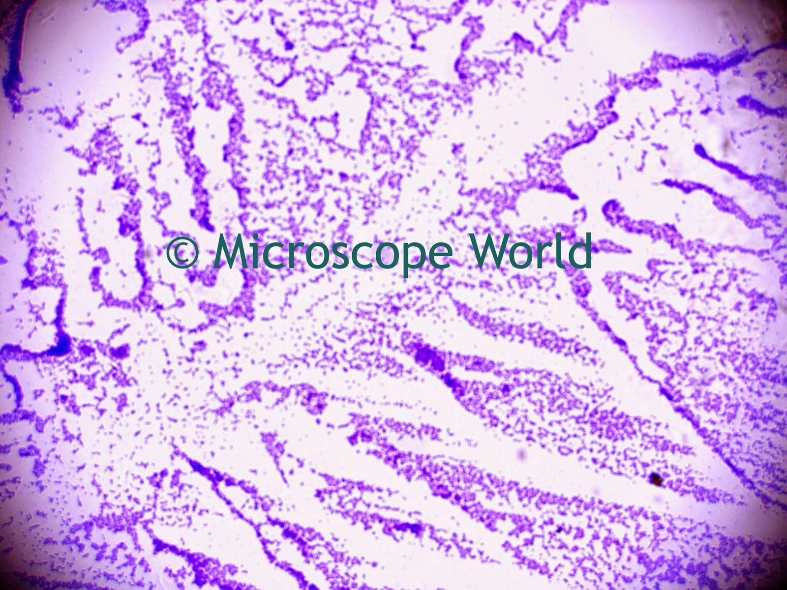
Microscope World Blog: Staphylococcus Under the Microscope

Staphylococcus epidermidis 1,000x 1 | Marc Perkins Photography

Characterization of S. epidermidis ATCC 12228. (A) Gram stain was... |  Download Scientific Diagram
Characterization of S. epidermidis ATCC 12228. (A) Gram stain was... | Download Scientific Diagram

Staphylococcus epidermidis - Wikipedia

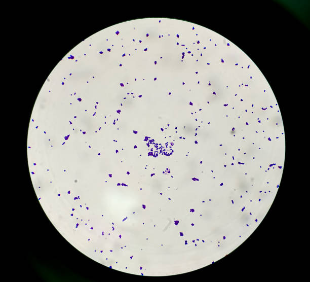
Staphylococcus epidermidis under microscope: microscopy of Gram-positive  cocci, morphology and microscopic appearance of Staphylococcus epidermidis,  S.epidermidis gram stain and colony morphology on agar, clinical  significance
Staphylococcus epidermidis under microscope: microscopy of Gram-positive cocci, morphology and microscopic appearance of Staphylococcus epidermidis, S.epidermidis gram stain and colony morphology on agar, clinical significance

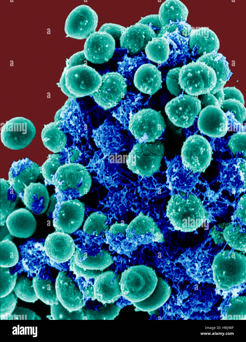
Staphylococcus epidermidis hi-res stock photography and images - Alamy